

Acute hemorrhagic encephalomyelitis in childhood: Case report and literature review
- PMID: 21977089
- PMCID: PMC3173916
- DOI: 10.4103/1817-1745.84408
Acute hemorrhagic encephalomyelitis in childhood: Case report and literature review
Abstract
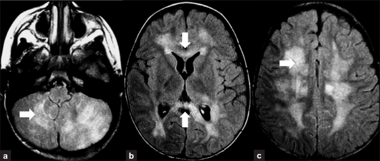
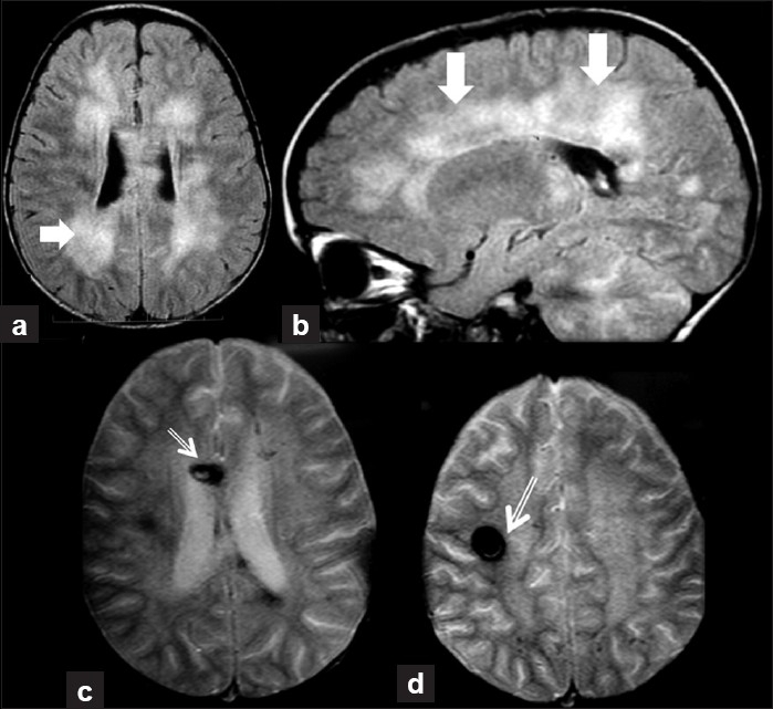
Acute disseminated encephalomyelitis (ADEM) is an inflammatory immune-mediated disorder which is more common in pediatric patients. The clinical setting is characterized by a rapid onset of encephalopathy and multifocal neurological features. Acute hemorrhagic encephalomyelitis (AHEM) is considered a rare form of ADEM. This report shows a 2-year-old patient who presented with the classical features of ADEM and after 8 weeks developed severe neurological worsening. The second magnetic resonance image (MRI) showed hemorrhagic lesions. Differences in prognosis between ADEM and AHEM justify the investigation of AHEM whenever a patient has neurological recrudescence in a known patient of ADEM.
Keywords: Acute disseminated encephalomyelitis; acute hemorrhagic; childhood.
Conflict of interest statement
Figures
References
-
- Tenembaum S, Chitnis T, Ness J, Hahn JS. International Pediatric MS Study Group. Acute disseminated encephalomyelitis. Neurology. 2007;68(16 Suppl 2):S23–36. - PubMed
-
- Dale RC, de Sousa C, Chong WK, Cox TC, Harding B, Neville BG. Acute disseminated encephalomyelitis, multiphasic disseminated encephalomyelitis and multiple sclerosis in children. Brain. 2000;123:2407–22. - PubMed
-
- Mikaeloff Y, Suissa S, Vallée L, Lubetzki C, Ponsot G, Confavreux C, et al. First episode of acute CNS inflammatory demyelination in childhood: Prognostic factors for multiple sclerosis and disability. J Pediatr. 2004;144:246–52. - PubMed
-
- Leake JA, Billman GF, Nespeca MP, Duthie SE, Dory CE, Meltzer HS, et al. Pediatric acute hemorrhagic leukoencephalitis: Report of a surviving patient and review. Clin Infect Dis. 2002;34:699–703. - PubMed
-
- Rosman NP, Gottlieb SM, Bernstein CA. Acute hemorrhagic leukoencephalitis: Recovery and reversal of magnetic resonance imaging findings in a child. J Child Neurol. 1997;12:448–54. - PubMed
