

Three-dimensional echocardiography in valve disease
- PMID: 21977273
- PMCID: PMC3184677
- DOI: 10.4081/hi.2007.35
Three-dimensional echocardiography in valve disease
Abstract
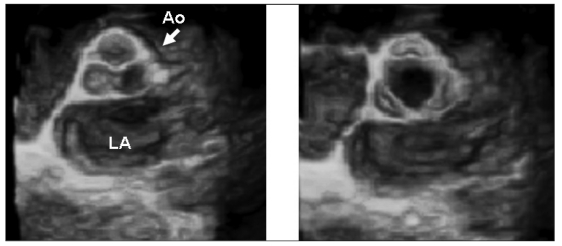
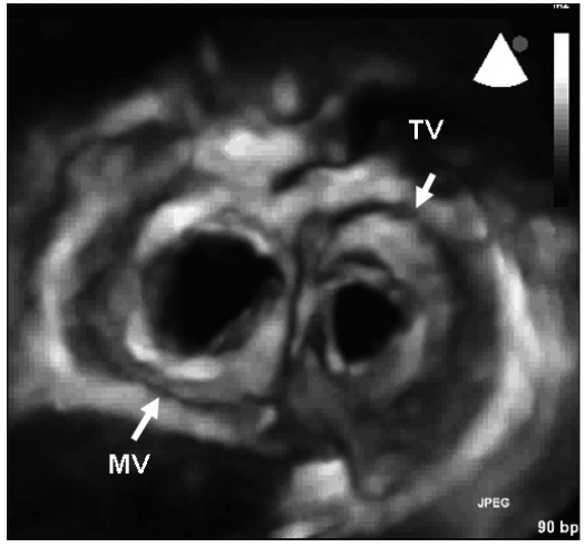
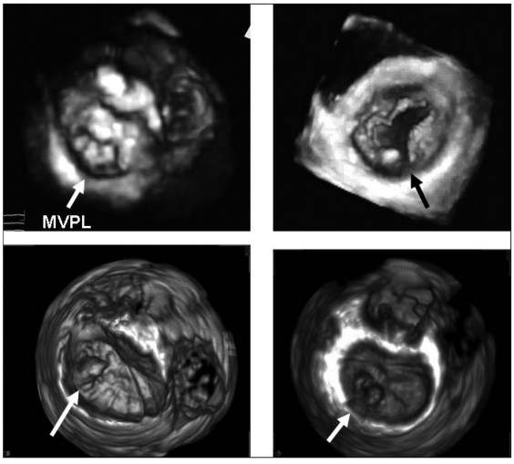
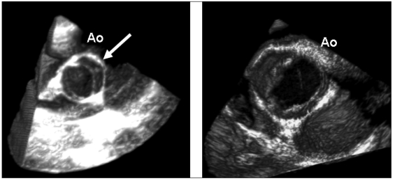
This review covers the role of three-dimensional (3D) echocardiography in the diagnosis of heart valve disease. Several factors have contributed to the evolution of this technique, which is currently a simple and routine method: rapid evolution in probe and computer technologies, demonstration that 3D data sets allowed more complete and accurate evaluation of cardiac structures, emerging clinical experience indicating the strong potential particularly in valve diseases, volume and function of the two ventricle measurements and several other fields. This report will review current and future applications of 3D echocardiography in mitral, aortic and tricuspid valve diseases underlying both qualitative (morphologic) and quantitative advantages of this technique.
Keywords: Three-dimensional transesophageal echocardiography; Three-dimensional transthoracic echocardiography; Valve disease.
Figures
Similar articles
-
Real-time three-dimensional transesophageal echocardiography--an initial experience.Rev Port Cardiol. 2009 Jun;28(6):671-82. Rev Port Cardiol. 2009. PMID: 19697795 English, Portuguese.
-
Dynamic three-dimensional echocardiography: a new era in ultrasound technology.Rev Port Cardiol. 1997 Oct;16(10):787-95, 745-6. Rev Port Cardiol. 1997. PMID: 9436415 Review.
-
Three-dimensional echocardiography for the assessment of the tricuspid valve.Echocardiography. 2020 May;37(5):758-768. doi: 10.1111/echo.14658. Epub 2020 Apr 21. Echocardiography. 2020. PMID: 32315483 Review.
-
Dynamic three-dimensional reconstruction of the heart by transesophageal echocardiography.Arq Bras Cardiol. 1999 May;72(5):559-68. doi: 10.1590/s0066-782x1999000500003. Arq Bras Cardiol. 1999. PMID: 10668226 English, Portuguese.
-
Initial experience with a new on-line transthoracic three-dimensional technique: assessment of feasibility and of diagnostic potential.Ital Heart J. 2003 Aug;4(8):544-50. Ital Heart J. 2003. PMID: 14564981 Clinical Trial.
References
-
- Badano LP, Dall’Armellina E, Monaghan MJ, et al. Real-time three-dimensional echocardiography: technological gadget or clinical tool? J Cardiovasc Med. 2007;8:144–62. - PubMed
-
- Roelandt JR, Yao J, Kasprzak JD. Three-dimensional echocardiography. Curr Opin Cardiol. 1998;13:386–96. - PubMed
-
- Levine RA, Weyman AE, Hand Shumacher MD. Three-dimensional echocardiography: techniques and applications. Am J Cardiol. 1992;69:121–34. - PubMed
-
- Kisslo J, Firek B, Ota T, et al. Real-time volumetric echocardiography: the technology and the possibilities. Echocardiography. 2000;17:773–9. - PubMed
-
- Pepi M, Tamborini G, Pontone G, et al. Initial experience with a new on-line transthoracic three-dimensional technique: assessment of feasibility and of diagnostic potential. Ital Heart J. 2003;4:544–50. - PubMed
LinkOut - more resources
Full Text Sources