Initial experience using contrast enhanced real-time three-dimensional exercise stress echocardiography in a low-risk population
- PMID: 21977293
- PMCID: PMC3184705
- DOI: 10.4081/hi.2010.e8
Initial experience using contrast enhanced real-time three-dimensional exercise stress echocardiography in a low-risk population
Abstract
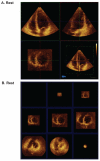
Although emerging data support the utility of real-time three-dimensional echocardiography (RT3DE) during dobutamine stress testing, the feasibility of performing contrast enhanced RT3DE during exercise treadmill stress has not been explored. Two-dimensional (2D) and three-dimensional (3D) acquisition were performed in 39 patients at rest and peak exercise. Contrast was used in 29 patients (74%). Reconstruction was performed manually by generating short axis cut planes at the base, mid-ventricle and apex, and automatically by generating 9 short axis slices. Three-dimensional acquisition was feasible during rest and stress regardless of the use of contrast. Time to acquire stress images was reduced using 3D (35.2±17.9 s) as compared to 2D acquisition (51.6±14.7 s; P<0.05). Using a 17-segment model, of all 663 segments, 588 resting (88.6%) and 563 stress segments (84.9%) were adequately visualized using manually reconstructed 3D data, compared with 618 resting (93.2%) and 606 stress segments (91.4%) using 2D data (P rest=0.06; P stress=0.07). We concluded that contrast enhanced RT3DE is feasible during treadmill stress echocardiography.
Keywords: three-dimensional exercise stress echocardiography..
Conflict of interest statement
Conflict of interest: the authors report no conflicts of interest.
Figures


Similar articles
-
Comparison of contrast-enhanced real-time live 3-dimensional dobutamine stress echocardiography with contrast 2-dimensional echocardiography for detecting stress-induced wall-motion abnormalities.J Am Soc Echocardiogr. 2006 Mar;19(3):294-9. doi: 10.1016/j.echo.2005.10.008. J Am Soc Echocardiogr. 2006. PMID: 16500492 Clinical Trial.
-
Non-invasive assessment of myocardial ischaemia using new real-time three-dimensional dobutamine stress echocardiography: comparison with conventional two-dimensional methods.Eur Heart J. 2005 Aug;26(16):1625-32. doi: 10.1093/eurheartj/ehi194. Epub 2005 Apr 7. Eur Heart J. 2005. PMID: 15817607
-
Usefulness of ultrasound contrast agent to improve image quality during real-time three-dimensional stress echocardiography.Am J Cardiol. 2007 Jan 15;99(2):275-8. doi: 10.1016/j.amjcard.2006.08.023. Epub 2006 Nov 27. Am J Cardiol. 2007. PMID: 17223433
-
Real time three-dimensional stress echocardiography advantages and limitations.Echocardiography. 2012 Feb;29(2):200-6. doi: 10.1111/j.1540-8175.2011.01626.x. Echocardiography. 2012. PMID: 22283201 Review.
-
Stress echocardiography using real-time three-dimensional imaging.Echocardiography. 2018 Aug;35(8):1196-1203. doi: 10.1111/echo.14050. Epub 2018 Jun 20. Echocardiography. 2018. PMID: 30133883 Review.
References
-
- Pellikka PA, Nagueh SF, Elhendy AA, et al. American society of echocardiography recommendations for performance, interpretation, and application of stress echocardiography. J Am Soc Echocardiogr. 2007;20:1021–41. - PubMed
-
- Armstrong WF, Zoghbi WA. Stress echocardiography: current methodology and clinical applications. J Am Coll Cardiol. 2005;45:1739–47. - PubMed
-
- Collins M, Hsieh A, Ohazama CJ, et al. Assessment of regional wall motion abnormalities with real-time three-dimensional echocardiography. J Am Soc Echocardiogr. 1999;12:7–14. - PubMed
-
- Hung J, Lang R, Flachskampf F, et al. 3D echocardiography: A review of current status and future directions. J Am Soc Echocardiogr. 2007;20:213–33. - PubMed
Publication types
LinkOut - more resources
Full Text Sources
Miscellaneous
