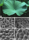
Superhydrophobicity in perfection: the outstanding properties of the lotus leaf
- PMID: 21977427
- PMCID: PMC3148040
- DOI: 10.3762/bjnano.2.19
Superhydrophobicity in perfection: the outstanding properties of the lotus leaf
Abstract
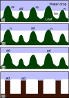
Lotus leaves have become an icon for superhydrophobicity and self-cleaning surfaces, and have led to the concept of the 'Lotus effect'. Although many other plants have superhydrophobic surfaces with almost similar contact angles, the lotus shows better stability and perfection of its water repellency. Here, we compare the relevant properties such as the micro- and nano-structure, the chemical composition of the waxes and the mechanical properties of lotus with its competitors. It soon becomes obvious that the upper epidermis of the lotus leaf has developed some unrivaled optimizations. The extraordinary shape and the density of the papillae are the basis for the extremely reduced contact area between surface and water drops. The exceptional dense layer of very small epicuticular wax tubules is a result of their unique chemical composition. The mechanical robustness of the papillae and the wax tubules reduce damage and are the basis for the perfection and durability of the water repellency. A reason for the optimization, particularly of the upper side of the lotus leaf, can be deduced from the fact that the stomata are located in the upper epidermis. Here, the impact of rain and contamination is higher than on the lower epidermis. The lotus plant has successfully developed an excellent protection for this delicate epistomatic surface of its leaves.
Keywords: Lotus effect; epicuticular wax; leaf surface; papillae; water repellency.
Figures


Similar articles
-
Superhydrophobic surfaces developed by mimicking hierarchical surface morphology of lotus leaf.Molecules. 2014 Apr 4;19(4):4256-83. doi: 10.3390/molecules19044256. Molecules. 2014. PMID: 24714190 Free PMC article. Review.
-
From natural to biomimetic: The superhydrophobicity and the contact time.Microsc Res Tech. 2016 Aug;79(8):712-20. doi: 10.1002/jemt.22689. Epub 2016 Jun 2. Microsc Res Tech. 2016. PMID: 27252147
-
Durable Bio-Based Hydrophobic Recrystallized Wax Coatings.ACS Appl Bio Mater. 2025 Feb 17;8(2):1437-1450. doi: 10.1021/acsabm.4c01672. Epub 2025 Jan 31. ACS Appl Bio Mater. 2025. PMID: 39887690
-
The role of bio-inspired hierarchical structures in wetting.Bioinspir Biomim. 2015 Apr 9;10(2):026009. doi: 10.1088/1748-3190/10/2/026009. Bioinspir Biomim. 2015. PMID: 25856043
-
A critical review on robust self-cleaning properties of lotus leaf.Soft Matter. 2023 Feb 8;19(6):1058-1075. doi: 10.1039/d2sm01521h. Soft Matter. 2023. PMID: 36637093 Review.
Cited by
-
Superhydrophobic hierarchically structured surfaces in biology: evolution, structural principles and biomimetic applications.Philos Trans A Math Phys Eng Sci. 2016 Aug 6;374(2073):20160191. doi: 10.1098/rsta.2016.0191. Philos Trans A Math Phys Eng Sci. 2016. PMID: 27354736 Free PMC article. Review.
-
Fabrication and Testing of Multi-Hierarchical Porous Scaffolds Designed for Bone Regeneration via Additive Manufacturing Processes.Polymers (Basel). 2022 Sep 27;14(19):4041. doi: 10.3390/polym14194041. Polymers (Basel). 2022. PMID: 36235989 Free PMC article.
-
Recent Advances in Surface Nanoengineering for Biofilm Prevention and Control. Part I: Molecular Basis of Biofilm Recalcitrance. Passive Anti-Biofouling Nanocoatings.Nanomaterials (Basel). 2020 Jun 24;10(6):1230. doi: 10.3390/nano10061230. Nanomaterials (Basel). 2020. PMID: 32599948 Free PMC article. Review.
-
Development of a Monitoring Strategy for Laser-Textured Metallic Surfaces Using a Diffractive Approach.Materials (Basel). 2019 Dec 20;13(1):53. doi: 10.3390/ma13010053. Materials (Basel). 2019. PMID: 31861907 Free PMC article.
-
Production of Hydrophobic Zein-Based Films Bioinspired by The Lotus Leaf Surface: Characterization and Bioactive Properties.Microorganisms. 2019 Aug 16;7(8):267. doi: 10.3390/microorganisms7080267. Microorganisms. 2019. PMID: 31426406 Free PMC article.
References
-
- Barthlott W. Die Selbstreinigungsfähigkeit pflanzlicher Oberflächen durch Epicuticularwachse. In: Rheinische Friedrich-Wilhelms-Universität Bonn., editor. Klima- und Umweltforschung an der Universität Bonn. Bonn: Bornemann; 1992. pp. 117–120.
-
- Barthlott W, Neinhuis C. Planta. 1997;202:1–8. doi: 10.1007/s004250050096. - DOI
-
- Barthlott W, Neinhuis C. Biol Unserer Zeit. 1998;28:314–321. doi: 10.1002/biuz.960280507. - DOI
-
- Bhushan B, Jung C J, Nosonovsky M. Lotus Effect: Surfaces with Roughness-Induced Superhydrophobicity, Self-Cleaning, and Low Adhesion. In: Bhushan B, editor. Springer Handbook of Nanotechnology. 3rd ed. New York: Springer; 2010. pp. 1437–1524.
LinkOut - more resources
Full Text Sources
Other Literature Sources
