. 2011 Nov;134(3):270-80.
doi: 10.1111/j.1365-2567.2011.03486.x.

Transient modification within a pool of CD4 T cells in the maternal spleen

Affiliations

Transient modification within a pool of CD4 T cells in the maternal spleen

Elizabeth A Bonney et al. Immunology. 2011 Nov.

Abstract

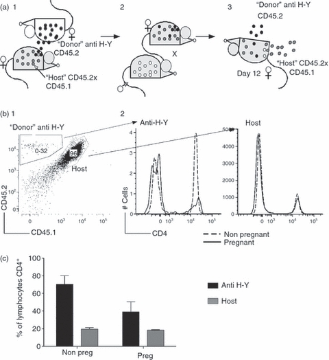
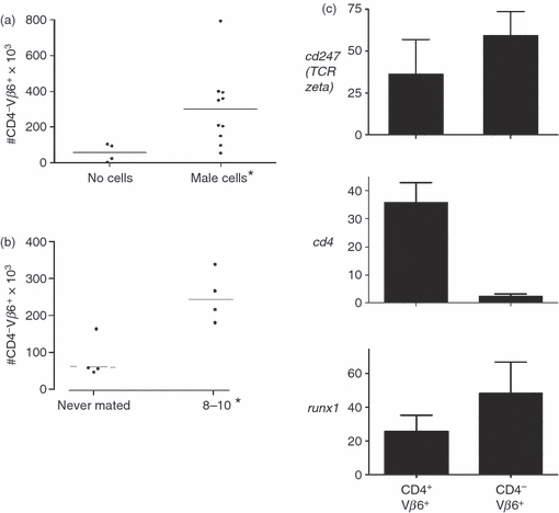
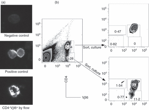
Classic models suggest maternal tolerance is dependent on regulation of fetal antigen-specific T cell responses. We hypothesize that factors unique to a particular fetal antigen-specific T cell, rather than the state of pregnancy per se, are important determinants of T cell fate during pregnancy. To investigate the fate of fetal antigen-specific CD4 T cells in the systemic circulation, we examined spleen cells in a CD4 T cell receptor transgenic mouse specific for the male antigen H-Y. We observed a transient decrease in CD4(+) Vβ6(+) cell numbers and, due to transient internalization of CD4, an increase in CD4(-) Vβ6(+) T cells. Antigen-specific in vitro responsiveness was not depressed by pregnancy. These data suggest that pregnancy supports fluidity in this particular CD4 T cell pool that may, in turn, help to meet competing requirements of maternal immune responsiveness and fetal tolerance.


Figures

Figure 1
Figure 1
Analysis of CD4 T cells during pregnancy in normal mice. C57BL/6 females were either left naive or mated with same-strain males and euthanized on various days of pregnancy to harvest, analyse and enumerate CD4+ T cells. (a) Example of flow cytometric analysis. Pink quadrant: CD25+ CD44low. Blue quadrant: CD44 intermediate CD25+. Yellow quadrant: CD44high CD25. (b) Comparison of spleen and uterine draining node CD4+ T cells. Upper panels: spleen. Lower panels: uterine draining nodes. y-Axis: percentage of CD4+ T cell receptor β (TCR-β)+ cells; x-axis: gestational age where ‘um’ denotes unmated mice. Pink bars: percentage of cells falling in pink quadrant by flow cytometry. Blue bars: percentage of cells falling in the blue quadrant. Yellow bars: percentage of cells falling in the yellow quadrant. Each bar represents the mean (± standard error of the mean) of at least five mice. Analysis of the data was performed by Kruskal–Wallis test followed by Dunn's multiple comparisons test. Asterisk: P < 0·05. (c) Enumeration of cells in the spleen. y-Axis: number of cells; x-axis: gestational age; ‘um’ denotes unmated mice. Each symbol represents one mouse. Left: CD4+ TCR-β+ cells. Right: CD4+ TCR+ CD44high CD25 cells. Analysis of the data was performed by Kruskal–Wallis test followed by Dunn's multiple comparisons test. Asterisk: P < 0·05.
Figure 2
Figure 2
Transient loss of CD4+ Vβ6+ spleen T cells during pregnancy in the spleen of CD4 anti H-Y transgenic mice. (a) Example of flow cytometric analysis. Spleen cells from a never mated (left panel), day 12 pregnant (middle panel) and a post-partum (right panel) mouse were analysed using antibodies to CD4 (y-axis) and Vβ6 (x-axis). (b) Comparison of spleen and uterine draining lymph node. Spleen (left panel, n = 5–8 mice each time-point) and uterine draining lymph nodes (right panel, n = 5–8 mice each time-point) were harvested from pregnant and unmated mice and analysed as in (a). y-Axis: percentage of lymphocytes positive for CD4 and Vβ6; x-axis: time of assay, where ‘um’ refers to unmated mice and ‘post-partum’ refers to mice < 2 weeks from birth of a litter. Asterisk: P < 0·05.
Figure 3
Figure 3
The spleen of CD4 anti-H-Y transgenic mice retains responsiveness despite transient decrease in numbers of CD4+ Vβ6+ spleen T cells during pregnancy. (a) Enumeration of CD4+ Vβ6+ spleen T cells. y-Axis: number of cells; x-axis: status; ‘um’ denotes unmated mice. Each symbol represents one mouse. Asterisk denotes a statistically significant (P < 0·05) difference compared to never-mated mice after application of the Kruskal–Wallis test. (b) Relative expression of T cell receptor zeta and CD4 in CD4+ Vβ6+ CD4 H-Y T cell receptor (TCR) transgenic spleen cells. CD4 H-Y TCR transgenic spleen cells from never mated (white bars, n = 3) and gestational age 12 (black bars, n = 3) were subjected to flow sorting, and the CD4+ Vβ6+ spleen cells (> ∼95% purity) were used to extract RNA which was then used to determine gene expression by quantitative reverse transcription–polymerase chain reaction (qRT–PCR). y-Axis: relative expression; x-axis: gene tested. Comparison of all groups by Kruskal–Wallis test and Dunn's post-test reveals P = 0·24. (c) CD4 H-Y TCR transgenic CD4+ Vβ6+ T cells retain the ability to respond to male cells. y-Axis: specific proliferation of 100 000 CD4 H-Y TCR transgenic spleen cells towards 500 000 male cells divided by the proliferation towards the same number of female spleen cells and by the percent of CD4+ Vβ6+ cells in the input culture as determined by flow cytometry in two experiments. Bars: median values of mice assayed. Numbers in brackets depict the range for the assay; x-axis: gestational age and number of mice assayed.
Figure 4
Figure 4
Decrease in the numbers of CD4-expressing cells in the spleen of anti-H-Y T cell receptor (TCR) transgenic mice during pregnancy is antigen-specific. (a) Course of experiment. (1) Normal CD45.2+ CD45.1+ females were injected with CD4 anti-H-Y TCR transgenic RAGKO spleen cells (CD45.2 single-positive, cells presented as black circles) as neonates and allowed to mature to create stable chimeras. (2) Stable chimeras were mated with CD45.1+ males (cells represented as white circles) or left naive. (3) At mid-gestation (day 10), pregnant mice were euthanized and spleens were analysed for the percentage of host (CD45.2+ CD45.1+, grey) or donor (anti H-Y, CD45.2 single-positive, black) lymphocytes expressing CD4. (b) Example of flow cytometry. Spleen cells falling in the lymphocyte gate (not shown) were first analysed for the presence of donor or host markers. These cells were analysed in turn for the surface expression of CD4. (1) Example of spleen from a pregnant chimeric mouse with CD4 H-Y TCR transgenic cells present (∼0·3%) and host cells (96%). A very small proportion of cells bearing only the CD45.1 marker may be of fetal origin. (2) Comparison of CD4 expression in chimeric CD4 H-Y TCR transgenic or host cells. Dotted lines: histograms from a never mated, non-pregnant mouse. Solid lines: histograms from a pregnant mouse. (c) Behaviour of chimeric CD4 anti-H-Y TCR transgenic cells in normal mice. Data are representative of three pregnant and three non-pregnant chimeras. Black bars: anti-H-Y T cells. Grey bars: host T cells. y-Axis: percentage of cell type (donor or host) lymphocytes that are CD4+; x-axis: pregnancy status. Data analysis was performed using two-way analysis of variance (anova), with P = 0·06.
Figure 5
Figure 5
Increase in the number of CD4 Vβ6+ cells in the spleen of pregnant H-Y T cell receptor (TCR) transgenic mice. (a) CD4 Vβ6+ cells in the spleen of non-pregnant mice injected with male cells. Never-mated female H-Y TCR transgenic mice were injected with male cells or not given cells. Ten days later, the mice were euthanized and their spleens harvested and analysed by flow cytometry. y-Axis: numbers CD4 Vβ6+ of cells; x-axis: treatment. Asterisk: P < 0·05. (b) CD4 Vβ6+ cells in the spleen of pregnant and non-pregnant mice. y-Axis: numbers of cells; x-axis: pregnancy status. Asterisk: P < 0·05. (c) Analysis of spleen CD4+ Vβ6+ (double-positive) and CD4 Vβ6+ spleen cells in pregnant mice. Female CD4 anti-H-Y TCR transgenic mice were mated and on day 12 of gestation they were euthanized and the spleens were harvested and subjected to flow sorting as in Fig. 2c. RNA was extracted from three pools each of flow-purified CD4+ Vβ6+ (double-positive) and CD4 Vβ6+ and this was used to determine the relative expression of Cd247 (top panel) Cd4 (middle panel, P = 0·057) and Runx1 (bottom panel). y-Axis: gene examined; x-axis: cell type.
Figure 6
Figure 6
CD4 down-regulation in the spleen cells of CD4 anti-H-Y T cell receptor (TCR) transgenic mice during pregnancy is transient. (a) Intracellular expression of CD4 on CD4 H-Y TCR transgenic spleen cells. CD4 H-Y TCR transgenic spleen cells that were identified as CD4 Vβ6+ or CD4+ Vβ6+ by flow cytometry were placed onto glass slides and analysed by microscopy to determine expression of CD4. Top: Representative cell on negative control slide. Middle: representative CD4+ Vβ6+ cell by flow cytometry. Bottom: representative CD4 Vβ6+ cells by flow cytometry. (b) Re-expression of CD4 in CD4 H-Y TCR transgenic spleen cells in pregnant mice. Left: initial sorting profile. Spleen cells from pooled CD4 H-Y TCR transgenic mice were stained with antibodies to CD4 and Vβ6 and sorted into subpools based on expression of the two markers. Each subpool was incubated in culture medium and then re-examined for expression of CD4 and Vβ6. Right: post-culture analysis of sorted cells. Top right: CD4+ Vβ6+ cells by initial flow cytometry. Bottom right: CD4− Vβ6+ cells by initial flow cytometry.

Similar articles

Cited by

References

    1. Munn DH, Zhou M, Attwood JT, Bondarev I, Conway SJ, Marshall B, Mellor AL. Prevention of allogeneic fetal rejection by tryptophan catabolism. Science. 1998;281:1191–3. - PubMed
    1. Zenclussen AC, Gerlof K, Zenclussen ML, et al. Regulatory T cells induce a privileged tolerant microenvironment at the fetal–maternal interface. Eur J Immunol. 2006;36:82–94. - PubMed
    1. Vacchio MS, Hodes RJ. Fetal expression of Fas ligand is necessary and sufficient for induction of CD8 T cell tolerance to the fetal antigen H-Y during pregnancy. J Immunol. 2005;174:4657–61. - PubMed
    1. Aluvihare VR, Kallikourdis M, Betz AG. Regulatory T cells mediate maternal tolerance to the fetus. Nat Immunol. 2004;5:266–71. - PubMed
    1. Bonney EA, Matzinger P. The maternal immune system's interaction with circulating fetal cells. J Immunol. 1997;158:40–7. - PubMed

Publication types

MeSH terms

LinkOut - more resources