. 2011 Nov 8;50(44):9520-31.
doi: 10.1021/bi2014695. Epub 2011 Oct 14.

Detergents stabilize the conformation of phosphodiesterase 6

Affiliations

Detergents stabilize the conformation of phosphodiesterase 6

Bo Y Baker et al. Biochemistry. .

Abstract

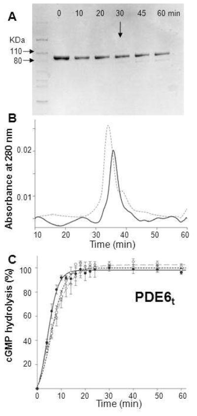
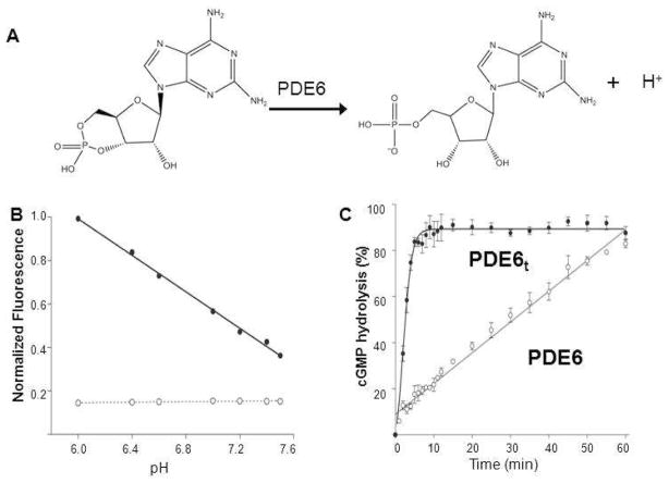
Membrane-bound phosphodiesterase 6 (PDE6) plays an important role in visual signal transduction by regulating cGMP levels in rod photoreceptor cells. Our understanding of PDE6 catalysis and structure suffers from inadequate characterization of the α and β subunit catalytic core, interactions of the core with two intrinsically disordered, proteolysis-prone inhibitory PDEγ (Pγ) subunits, and binding of two types of isoprenyl-binding protein δ, called PrBP/δ, to the isoprenylated C-termini of the catalytic core. Structural studies of native PDE6 have been also been hampered by the lack of a heterologous expression system for the holoenzyme. In this work, we purified PDE6 in the presence of PrBP/δ and screened for additives and detergents that selectively suppress PDE6 basal activity while sparing that of the trypsin-activated enzyme. Some detergents removed PrBP/δ from the PDE complex, separating it from the holoenzyme after PDE6 purification. Additionally, selected detergents also significantly reduced the level of dissociation of PDE6 subunits, increasing their homogeneity and stabilizing the holoenzyme by substituting for its native membrane environment.


Figures

Figure 1
Figure 1
Preparation of trypsinized PDE6. A. The time course of trypsin digestion monitored by SDS–PAGE is shown. Purified PDE6 was mixed with trypsin at 100:1 (weight ratio) and incubated at room temperature for 10, 20, 30, 45 and 60 min. Protein without trypsin was included as a control (Lane 0). At 30 min (arrow), a weak band with a smaller molecular weight appeared. B. Size exclusion chromatography profile of PDE6t (30 min, room temperature). PDE6t (solid line) eluted at nearly the same position as untreated PDE6 (dashed line). C. PDE6t activity with cGMP (2 mM) as substrate. Enzyme activities monitored with the pH–sensitive fluorescent dye SNARF–1were positively correlated with increasing fluorescence intensity over time. PDE6t was generated as follows: Purified PDE6 was first mixed with trypsin at a 100:1 weight ratio for 10 min. Soybean trypsin inhibitor was then added at a 1:10 mass ratio to stop the reaction (dashed line, –○–). Alternatively, PDE6 was mixed with trypsin–agarose at 30 μg protein per 1 μl agarose for 30 min and the reaction was stopped by passing the sample through a filter to remove the trypsin–agarose. Trypsinized peptides then were removed either by gel filtration (dashed line, –◇–) or buffer exchange with a 50 kDa MWCO filter (Millipore) (solid line, –•–). Activities for PDE6t prepared by all three methods were identical.
Figure 2
Figure 2
PDE6 activity measurements with the pH-sensitive fluorescent dye SNARF-1. A. Diagram presenting PDE6-catalyzed hydrolysis of cyclic GMP to GMP with release of a proton. B. PDE6 activity measurements with the pH-sensitive fluorescent dye SNARF-1 in 0.1 M HEPES over a pH range from 6.8 to 8.2. The fluorescence intensity of SNARF-1 decreased with increasing pH (–•–) whereas the buffer used showed no fluorescence (–○–). C. Enzyme activity measurements of PDE6 and PDE6t with SNARF-1. PDE6t achieved maximal cGMP hydrolysis in about 5 min (–•–), whereas PDE6 exhibited much lower residual activity that resulted in total cGMP hydrolysis in about 60 min (-○-). The percentage of cGMP hydrolysis is plotted as a function of time.
Figure 3
Figure 3
Effects of additives and detergents on PDE6t and PDE6 activity. A. Effects of reagents on PDE6t and PDE6 activity. Activities of purified PDE6t (upper panel) and PDE6 (lower panel) were measured in the absence (solid lines) or in the presence (dashed lines) of designated reagents. Urea (1 M, –▲–), Arginine (3 mM, –◆–), CaCl2 (0.1M, –X–) and EDTA (5 mM, –◇–). The percentage of maximum PDE6t activity vs. time is shown. B. Effects of selected detergents on PDE6 activity. The activities of PDE6t (upper panel) were measured in the absence (solid line) or in the presence of specified detergents (dashed lines). C8E4 (10 mM, –○–), Anapoe X–100 (0.8 mM, –◇–), CHAPS (25 mM, –Δ–), n–octyl–β–D–glucopyranoside (20 mM, –◆–), n–dodecyl–N,N–dimethylamine–N-oxide (1 mM, – formula image
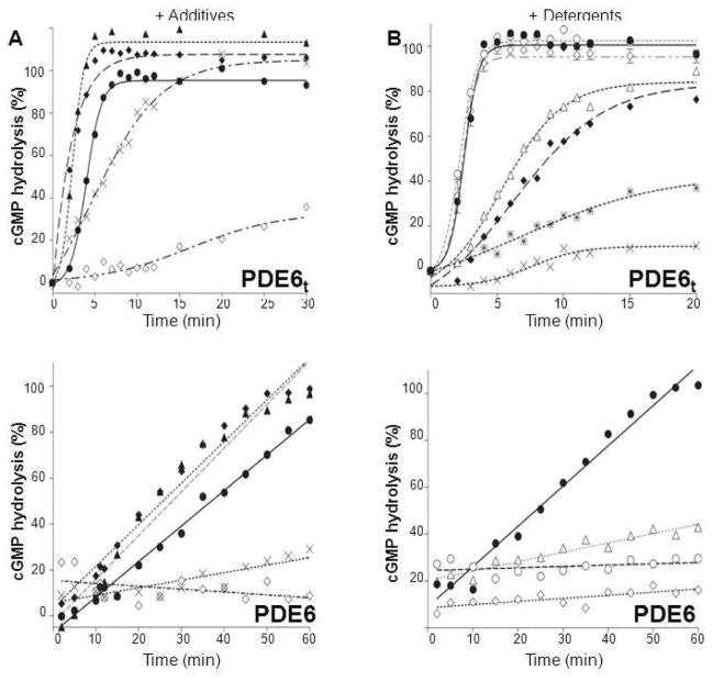
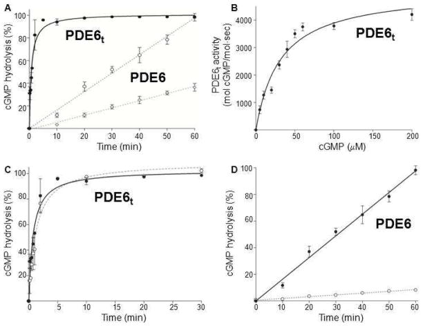
–) and dimethyldecylphosphine oxide (5 mM, –×–). Those detergents that did not alter PDE6t activity were then tested for their ability to suppress PDE6 activity (lower panel). PDE6 without detergent (solid line), with detergents (dashed lines). N–undecyl–β–D–maltopyranoside (1 mM, –Δ–), C8E4 (10 mM, –○–), Anapoe X–100 (0.8 mM, –◇–). The percentage of cGMP hydrolyzed is plotted vs. time.
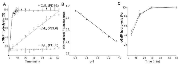
Figure 4
Figure 4
Effect of C8E4 on PDE6 activity. A. Measurements of PDE6t (bold black line, –•–) and PDE6 (dashed line, –○–) activity in the presence of C8E4. PDE6 activity without C8E4 is shown as a solid grey line (–○–). In the presence of C8E4, PDE6t was fully functional, whereas PDE6 activity was completely abolished. B. In the presence of C8E4, the fluorescence intensity of SNARF–1decreased with increasing pH, indicating that the dye responded appropriately to changes in pH. C. PDE6t activity in the presence of C8E4. Protein was pre–incubated in the assay buffer with C8E4 for 10 min before PDE6t activity was monitored (solid line). Alternatively, PDE6 was digested with trypsin in the presence of C8E4 before soybean trypsin inhibitor was added to stop the protease reaction and the resulting PDE6t was added to assay buffer along with C8E4 for activity measurements (dashed line). Both preparations of PDE6t were fully active.
Figure 5
Figure 5
SEC analysis of detergent effects on reducing PG aggregation. A. Elution profiles of three forms of PDE6 in the absence of detergent. PDE6 (short dashed line) and PDE6/δ (solid line) eluted as single peaks, whereas PG (large dashed line) eluted as two major peaks indicating a mixture of a high molecular mass aggregate and a monomer. Insert: SDS–PAGE analysis of SEC fractions from the two PG peaks. The top band is PDE6 and lower band is GST–δ. Some detergents, such as Anapoe–35 (B), reduced aggregation of PG. Insert: SDS–PAGE analysis of SEC fractions (indicated with dotted line). In the presence of C8E4 (C), PG aggregation was almost completely abolished. PDE6 was found mainly in Peak 1 (insert, labeled as 1). The second major peak eluting after the PDE6 peak represented GST– PrBP/δ as indicated by SDS–PAGE analysis (insert, labeled as 2). D. SEC analysis of the PDE6/δ conformation in the presence of C8E4; 0.5 ml fractions were collected SDS–PAGE of fractions from the two major peaks is shown in the insert. The left band (labeled with 1) shows the fraction from the main peak with an estimated MW of 200 kDa whereas the right band illustrates the fraction from the last peak with an estimated MW of 15 kDa (as indicated with an arrow). For all inserts, analysis of the protein sample before SEC is shown next to the protein marker. The top band corresponded to PDE6 and lower band to GST–δ.
Figure 6
Figure 6
SDS–PAGE analysis of detergent–depleted GST–PrBP/δ dissociated from the PG complex. A. C8E4 removed GST–PrBP/δ from the PG complex. PG was mixed with GST–FF resin and unbound proteins were removed by washing the resin with PBS. Then the resin was incubated with PBS plus 10 mM C8E4 for 1 hr at room temperature. The flow through was collected (Lane F, under C8E4) and the resin was washed again with PBS plus C8E4. Elution was accomplished with reduced glutathione (10 mM). F: flow through; W: wash fractions; E: Elution fraction. B. Dissociation of the PG complex was decreased by incubation with CnEm detergents with longer alkyl chains. CnEm: Cn = CH3(CH2)n–1 alkyl chain, Em = (OCH2CH2)mOH oligoethyleneglycol. In the presence of C8E4 or C8E6, PDE6 was identified mainly in SEC flow through fractions as shown by SDS–PAGE. But in the presence of C12E8, the PG complex remained intact in the elution fraction. Lanes F and E: flow through and elution fractions in the presence of CnEm.
Figure 7
Figure 7
Catalytic properties of purified PDE6t and PDE6 assessed directly by product formation. A. Percentages of cGMP hydrolysis by PDE6t and PDE6 are plotted as a function of time. PDE6t: solid line. PDE6: dashed lines (2 mM, –○– and 5 nM–◇–). B. The rate of cGMP hydrolysis by PDE6t is plotted as a function of cGMP concentration. The Km value is 31 ± 4 μM and Vmax is 5122 ± 285 mol cGMP/mol·sec. C: PDE6t activity in the presence and absence of C8E4. The percentage of cGMP hydrolysis by PDE6t is plotted as a function of time. PDE6t without C8E4: solid line. PDE6t with C8E4: dashed line. D. cGMP hydrolysis in the presence and absence of C8E4. The percentage of cGMP hydrolysis by PDE6 is plotted as a function of time. PDE6 without C8E4: solid line. PDE6 with C8E4: dashed line.
Figure 8
Figure 8
EM images of negatively stained PDE6/δ complexes in the absence and presence of indicated detergents. The protein retained homogeneity in all detergents tested except CHAPS in which the complex seems to have dissociated into particles of smaller size. Inset, higher magnification of a single particle. Scale bars, 100 nm.

Similar articles

Cited by

References

    1. Polans A, Baehr W, Palczewski K. Turned on by Ca2+! The physiology and pathology of Ca(2+)-binding proteins in the retina. Trends Neurosci. 1996;19:547–554. - PubMed
    1. Conti M, Beavo J. Biochemistry and physiology of cyclic nucleotide phosphodiesterases: essential components in cyclic nucleotide signaling. Annu Rev Biochem. 2007;76:481–511. - PubMed
    1. Artemyev NO, Surendran R, Lee JC, Hamm HE. Subunit structure of rod cGMP-phosphodiesterase. J Biol Chem. 1996;271:25382–25388. - PubMed
    1. Baehr W, Devlin MJ, Applebury ML. Isolation and characterization of cGMP phosphodiesterase from bovine rod outer segments. J Biol Chem. 1979;254:11669–11677. - PubMed
    1. Deterre P, Bigay J, Forquet F, Robert M, Chabre M. cGMP phosphodiesterase of retinal rods is regulated by two inhibitory subunits. Proc Natl Acad Sci U S A. 1988;85:2424–2428. - PMC - PubMed

Publication types

MeSH terms

LinkOut - more resources