

Dynamic CT technique for assessment of wrist joint instabilities
- PMID: 21978117
- PMCID: PMC3616456
- DOI: 10.1118/1.3577759
Dynamic CT technique for assessment of wrist joint instabilities
Abstract
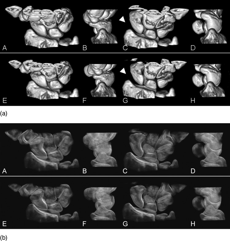
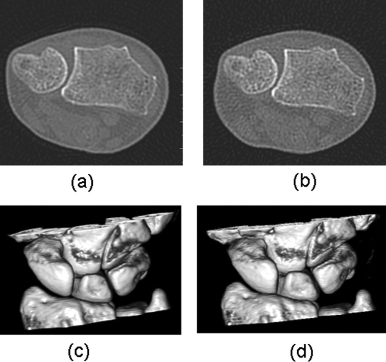
Purpose: To develop a 4D [three-dimensional (3D) + time] CT technique to capture high spatial and temporal resolution images of wrist joint motion so that dynamic joint instabilities can be detected before the development of static joint instability and onset of osteoarthritis (OA).
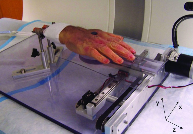
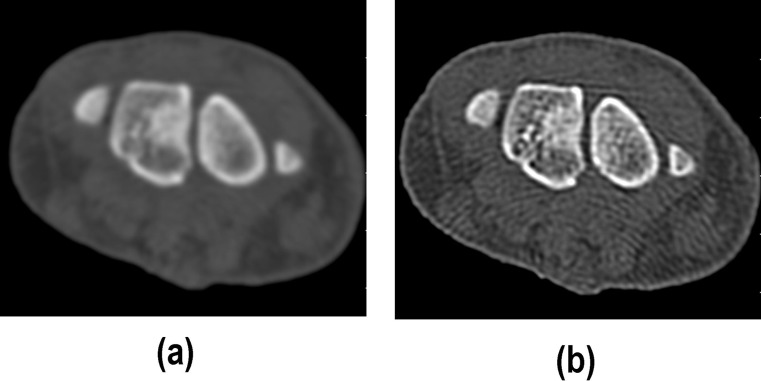
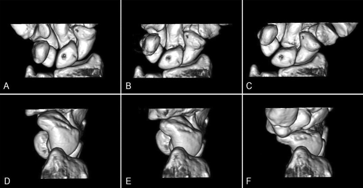
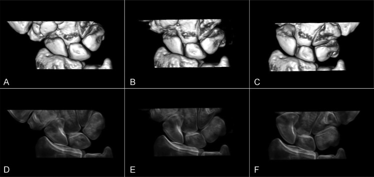
Methods: A cadaveric wrist was mounted onto a custom motion simulator and scanned with a dual source CT scanner during radial-ulnar deviation. A dynamic 4D CT technique was utilized to reconstruct images at 20 equidistant time points from one motion cycle. 3D images of carpal bones were generated using volume rendering techniques (VRT) at each of the 20 time points and then 4D movies were generated to depict the dynamic joint motion. The same cadaveric wrist was also scanned after cutting all portions of the scapholunate interosseus ligament to simulate scapholunate joint instability. Image quality were assessed on an ordinal scale (1-4, 4 being excellent) by three experienced orthopedic surgeons (specialized in hand surgery) by scoring 2D axial images. Dynamic instability was evaluated by the same surgeons by comparing the two 4D movies of joint motion. Finally, dose reduction was investigated using the cadaveric wrist by scanning at different dose levels to determine the lowest radiation dose that did not substantially alter diagnostic image quality.
Results: The mean image quality scores for dynamic and static CT images were 3.7 and 4.0, respectively. The carpal bones, distal radius and ulna, and joint spaces were clearly delineated in the 3D VRT images, without motion blurring or banding artifacts, at all time points during the motion cycle. Appropriate viewing angles could be interactively selected to view any articulating structure using different 3D processing techniques. The motion of each carpal bone and the relative motion among the carpal bones were easily observed in the 4D movies. Joint instability was correctly and easily detected in the scan performed after the ligament was cut by observing the relative motion between the scaphoid and lunate bones. Diagnostic capability was not sacrificed with a volume CT dose index (CTDI(vol)) as low as 18 mGy for the whole scan, with estimated skin dose of approximately 33 mGy, which is much lower than the threshold for transient skin erythema (2000 mGy).
Conclusions: The proposed dynamic 4D CT imaging technique generated high spatial and high temporal resolution images without requiring periodic joint motion. Preliminary results from this cadaveric study demonstrate the feasibility of detecting joint instability using this technique.
Figures
References
-
- Yelin E., “The economics of osteoarthritis,” (Osteoarthritis, Oxford University Press, New York, 1998), pp. 23–30.
Publication types
MeSH terms
Grants and funding
LinkOut - more resources
Full Text Sources
