Review
. 2012 Apr;36(2):122-9.
doi: 10.1016/j.clinre.2011.08.010. Epub 2011 Oct 5.

New pathophysiological concepts underlying pathogenesis of pigment gallstones

Affiliations
Review

New pathophysiological concepts underlying pathogenesis of pigment gallstones

Libor Vítek et al. Clin Res Hepatol Gastroenterol. 2012 Apr.

Abstract

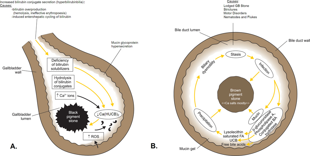
Pigment gallstones, which are much less frequent than cholesterol stones, are classified descriptively as "black" or "brown". They are composed mostly of calcium hydrogen bilirubinate, Ca(HUCB)(2), which is polymerized and oxidized in "black" stones but remains unpolymerized in "brown" stones. Black stones form in sterile gallbladder bile but brown stones form secondary to stasis and anaerobic bacterial infection in any part of the biliary tree, including the gallbladder. Other calcium salts coprecipitate in both stone types; crystalline calcium phosphate and/or carbonate in the case of "black" stones and amorphous calcium salts of long chain saturated fatty acids ("soaps") in the case of "brown" stones. Cholesterol is present in variable proportions in "brown" more than "black" stones and in the latter, the bile sterol may be totally absent. The "scaffolding" of both stone types is a mixed mucin glycoprotein matrix secreted by epithelial cells lining the biliary tree. The critical pathophysiological prerequisite for "black" stone formation is "hyperbilirubinbilia" (biliary hypersecretion of bilirubin conjugates). It is due principally to hemolysis, ineffective erythropoiesis, or pathologic enterohepatic cycling of unconjugated bilirubin. Endogenous biliary β-glucuronidase hydrolysis of bilirubin conjugates in gallbladder bile provides HUCB(-) molecules that precipitate as insoluble salts with ionized Ca. Putatively, reactive oxygen species secreted by an inflamed gallbladder mucosa are responsible for transforming the initial soft yellow precipitates into hard black [Ca(HUCB)(2)](n) polymers. Despite "brown" gallstones being soft and amenable to mechanical removal, chronic anaerobic infection of the biliary tree is often markedly resistant to eradication.


Figures

Fig 1
Fig 1
Schematic outline of the pathogenesis of: A, ‘black’ pigment stones in sterile gallbladder bile and B, ‘brown’ pigment stones in an obstructed biliary tree (gallbladder infrequently) infected with a mixed anaerobic microflora derived from the colon. In A, increased HUCB levels in bile arise from endogenous acidic β-glucuronidase, principally of hepatic origin, that hydrolyzes excess bilirubin glucuronides in bile (hyperbilirubinbilia) to UCB. Deficiency of solubilizers for Ca2+, monoacidic unconjugated bilirubin (HUCB), and Ca bilirubinates (bile salt-cholesterol micelles, bile salt-lecithin-cholesterol micelles, and unilamellar lecithin-cholesterol vesicles) leads to increases in free Ca2+ ions and free bilirubinate anions. Likewise, increases in free Ca2+ ions may be due to increased secretion into the biliary tree. In slightly alkaline bile, the concentrations of other Ca2+-sensitive anions, such as carbonate and phosphate, may also increase. When the ion products of the Ca-anion salts exceed their equilibrium solubility products in bile, the biliary system becomes supersaturated and precipitation is thermodynamically possible. Mucin glycoproteins are hypersecreted by the gallbladder mucosa like in other lithogenic settings and form a biliary gel providing a nucleation matrix for the precipitated pigment salts. Cholesterol may or may not phase-separate depending on its level in bile and is absent in stones when the cholesterol saturation index (CSI) of bile is less than 1, as occurs in most black pigment stone gallbladder biles. Solid-state ‘attack’ by free radicals or singlet oxygen (reactive oxygen species, ROS), from an inflamed gallbladder mucosa leads to tetrapyrrole polymerization and oxidation, which imparts the black spiculated appearance to most of these stones. In B, stasis, due to motor disorders of the sphincter of Oddi, strictures from biliary surgery or foreign bodies such as gallstones from the gallbladder, sutures, parasites and their eggs and carcasses (principally from nematodes and flukes), facilitate anaerobic bacterial invasion and overgrowth. Lithogenesis, stasis and infection lead to hypersecretion of biliary tree mucin glycoproteins that form a gel in bile. Bacterial enzymes catalyze the hydrolysis of ester and amide linkage in all biliary lipids: Phospholipase A1 catalyzes hydrolysis of biliary phosphatidylcholines at the sn-1 position to produce palmitic and stearic acids plus lysophosphatidylcholine; β-glucuronidase catalyzes hydrolysis of bilirubin glucuronosides to produce UCB and glucuronic acid which acidifies bile further; conjugated bile salt hydrolase (cholylglycylamidase) catalyzes hydrolysis of the amide linkage of conjugated bile salts to produce free (unconjugated) bile acids, taurine, or glycine. Saturated fatty acids can precipitate per se but typically form insoluble calcium soaps with Ca2+ ions; UCB forms insoluble acidic calcium bilirubinate salts Ca(HUCB)2, and free secondary bile acids may precipitate as such, or less commonly as calcium salts. Lysophosphatidylcholine may act as a fusogen for cholesterol-containing unilamellar phospholipid vesicles, leading to nucleation of cholesterol monohydrate crystals that are invariably co-precipitated with brown stones. The calcium salts, cholesterol, and biliary tree mucins, the major components of ‘brown’ pigment stones, also act as a ‘trap’ for the anaerobic bacteria, making their elimination difficult; bacterial ‘skeletons’ are usually visualized by electron microscopy of thick-sections of these stones. Ductal obstruction by brown stones themselves perpetuates the vicious cycle of stasis and chronic anaerobic bacterial infection. (Figure and legends A/B reproduced in modified form from reference 2 with written permission of the copyright owner: Thieme Medical Publishers, Inc. New York and Stuttgart, and redrawn by Mr. Martin Vyhnanovský, Prague, Czech Republic, and Ms. Jessica Y. Xia, Boston, MA, USA).

Comment in

References

    1. Schafmayer C, Hartleb J, Tepel J, Albers S, Freitag S, Völzke H, et al. Predictors of gallstone composition in 1025 symptomatic gallstones from Northern Germany. BMC Gastroenterol. 2006;6:36–44. - PMC - PubMed
    1. Cahalane MJ, Neubrand MW, Carey MC. Physical-chemical pathogenesis of pigment gallstones. Semin Liver Dis. 1988;8:317–328. - PubMed
    1. Suzuki N, Nakamura Y, Kobayashi N, Sato T. On metal elements in pure pigment gallstones. Tohoku J Exp Med. 1975;116:233–240. - PubMed
    1. Tsunoda K, Shirai Y, Wakai T, Yokoyama N, Akazawa K, Hatakeyama K. Increased risk of cholelithiasis after esophagectomy. J Hepatobiliary Pancreat Surg. 2004;11:319–323. - PubMed
    1. Strauss KA, Robinson DL, Vreman HJ, Puffenberger EG, Hart G, Morton DH. Management of hyperbilirubinemia and prevention of kernicterus in 20 patients with Crigler-Najjar disease. Eur J Pediatr. 2006;165:306–319. - PubMed

Publication types