. 2011 Oct 6:9:136.
doi: 10.1186/1477-7827-9-136.

Embryotoxicity assays for leached components from dental restorative materials

Affiliations

Embryotoxicity assays for leached components from dental restorative materials

Antonio Libonati et al. Reprod Biol Endocrinol. .

Abstract

Background: Currently, there are no suitable assays available to evaluate the embryotoxicity of leached components from restorative dental materials.

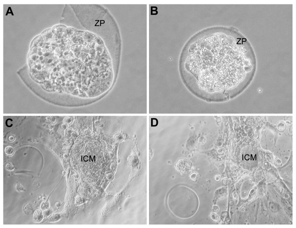
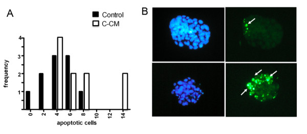
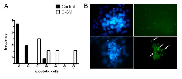
Methods: The effect of the medium conditioned by composites and amalgam on mouse blastocysts in vitro was tested. The materials were also subcutaneously implanted, and the effect of the medium supplemented with serum from the host blood was evaluated in the embryotoxicity assay. The embryo implantation rate in the material-transplanted mothers was also evaluated.

Results: The results show that while the culture in media conditioned by amalgams did not affect blastocyst development, the medium conditioned by composites caused blastocyst degeneration and apoptosis. The development of blastocysts in a medium containing serum obtained from animals after transplantation was, however, without effect. Finally, inconsistent reduction in the implantation rate in transplanted mothers was observed.

Conclusions: In this study, we provide examples of in vitro and in vivo tests that may be used to evaluate embryotoxicity for dental materials. Our results show that leached components from our composite-material induced embryotoxicity in vitro, however, no toxicity was observed when subcutaneously implanted in vivo. This highlights the necessity of integrated in vitro and in vivo tests for valuable predictive estimation of embryotoxicity for complex materials.


Figures

Figure 1
Figure 1
Morphologies and development of blastocysts in culture. Morphologies of blastocysts showing normal (A) or degenerated appearance (B) under phase contrast microscope. Note that blastocyst in A is undergoing zona pellucida (ZP) hatching. Normal inner cell mass (ICM) and trophoblast outgrowing (C) scored as +++, when compared with reduced tissue outgrowing (D) occurring in composite-conditioned medium and scored as ++. Magnification approximately 200 × (A, B) and 100 × (C, D).
Figure 2
Figure 2
Hoechst and TUNEL staining of blastocysts not capable of zona pellucida (ZP) hatching. A) Frequency histogram of non-ZP hatched blastocysts with different number of apoptotic cells evaluated by the nuclear staining, Hoechst 33342, cultured in control or composite-conditioned medium (C-CM) in three independent experiments. B) Pictures of representative blastocysts cultured in control (upper) or composite-conditioned medium (below) stained with Hoechst 33342 (left) and TUNEL method (right). The arrows indicate TUNEL-positive apoptotic cells. Note a few TUNEL-positive cells in the ICM of a control blastocyst and several TUNEL-positive cells mostly concentrated in the ICM of a blastocyst cultured in C-CM. Magnification approximately 100×.
Figure 3
Figure 3
Hoechst and TUNEL staining of blastocyst inner cell mass (ICM). Frequency histogram of ICM with different number of apoptotic cells evaluated by the nuclear staining, Hoechst 33342, of the embryo outgrowth cultured in control or composite-conditioned medium (C-CM) in three independent experiments. B) Pictures of representative ICM and trophoblast outgrowth cultured in control (upper) or composite-conditioned medium (below) stained with Hoechst 33342 (left) and by TUNEL method (right). The arrows indicate TUNEL-positive apoptotic cells. Note several TUNEL-positive cells in the ICM of a blastocyst cultured in C-CM. Magnification approximately 100×. All parameters were evaluated by observation under a phase contrast microscope as reported in the tests and showed in Figures 1, 2, 3. The results were obtained in at least three different experiments.

References

    1. Geurtsen W. Biocompatibility of resin-modified filling materials. Crit Rev Oral Biol Med. 2000;3:333–355. - PubMed
    1. Mackert JR. Dental amalgam and mercury. J Am Dent Assoc. 1991;122:54–61. - PubMed
    1. Reichl FX, Durner J, Muckter H, Elsenhans B, Forth W, Kunzelmann KH, Hickel R, Spahl W, Hume WR, Moes GW. Effect of dental materials on gluconeogenesis in rat kidney tubules. Arch Toxicol Suppl. 1999;73:381–386. doi: 10.1007/s002040050677. - DOI - PubMed
    1. Gerzina TM, Hume WR. Effect of dentine on release of TEGDMA from resin composite in vitro. J Oral Rehab. 1991;21:463–468. - PubMed
    1. Ferracane JL. Evolution of leach able components from composites. J Oral Rehab. 1994;21:441–452. doi: 10.1111/j.1365-2842.1994.tb01158.x. - DOI - PubMed

MeSH terms

LinkOut - more resources