. 2011 Oct 6:9:22.
doi: 10.1186/1478-811X-9-22.

RACK1, A multifaceted scaffolding protein: Structure and function

Affiliations

RACK1, A multifaceted scaffolding protein: Structure and function

David R Adams et al. Cell Commun Signal. .

Abstract

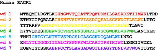
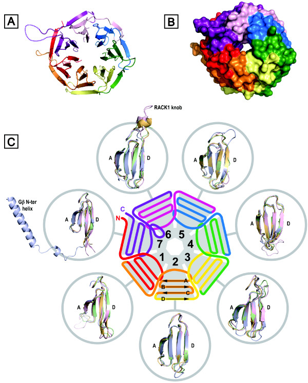
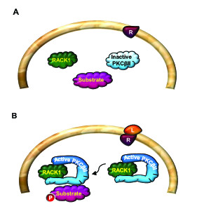
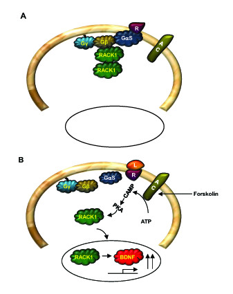
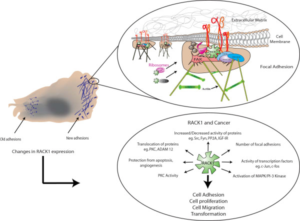
The Receptor for Activated C Kinase 1 (RACK1) is a member of the tryptophan-aspartate repeat (WD-repeat) family of proteins and shares significant homology to the β subunit of G-proteins (Gβ). RACK1 adopts a seven-bladed β-propeller structure which facilitates protein binding. RACK1 has a significant role to play in shuttling proteins around the cell, anchoring proteins at particular locations and in stabilising protein activity. It interacts with the ribosomal machinery, with several cell surface receptors and with proteins in the nucleus. As a result, RACK1 is a key mediator of various pathways and contributes to numerous aspects of cellular function. Here, we discuss RACK1 gene and structure and its role in specific signaling pathways, and address how posttranslational modifications facilitate subcellular location and translocation of RACK1. This review condenses several recent studies suggesting a role for RACK1 in physiological processes such as development, cell migration, central nervous system (CN) function and circadian rhythm as well as reviewing the role of RACK1 in disease.


Figures

Figure 1
Figure 1
Sequence of RACK1 depicting WD-repeats. RACK1 is a 317 amino acid protein with seven 40 amino acid repeats which are defined by sequences flanked by GH and WD dipeptides.
Figure 2
Figure 2
Structural detail for RACK1 proteins. (A) Crystal structure of RACK1A from A. thaliana (PDB: 3DM0), illustrating the seven-bladed β-propeller structure. (B) As (A) but with surface rendition. (C) Schematic representation of RACK1 structure and organisation of WD-repeats (defined in Figure 6); peripheral circles show superimposition of individual propeller blades for Gβ1 (blue) (PDB: 1TBG) and for the structurally defined RACK1 orthologues from A. thaliana (green) (PDB: 3DM0), S. cerevisiae (orange) (PDB: 3FRX) and T. thermophila (pink) (PDB: 2XZN). Gβ is distinguished in blade 7 by a helical N-terminal extension that engages tightly bound Gγ (not shown) in coiled coil interactions.
Figure 3
Figure 3
Model for PKCβII and RACK1 interaction. (A). In resting state, there is no interaction between RACK1 and inactive PKCβII. (B). Activation of PKCβII leads to the interaction between the active form of the kinase and RACK1. RACK1 then shuttles the active PKCβII to the site of its substrate.
Figure 4
Figure 4
Model for cAMP/PKA-mediated nuclear translocation of RACK1. (A). In resting state, RACK1 forms homodimers as well as heterodimers with Gβ. (B). Activation of the cAMP/PKA pathway leads to the dissociation of RACK1 from the complex and its translocation to the nucleus. In the nucleus RACK1 contributes to cAMP/PKA mediated increase in BDNF transcription.
Figure 5
Figure 5
Differential compartmentalization of RACK1 in the brain. (A). In hippocampal and dorsal striatal neurons, RACK1 is associated with both the NR2B subunit of the NMDAR and Fyn kinase. (B). cAMP/PKA activation or alcohol exposure leads to the release of RACK1 from the complex, enabling Fyn to phoshorylate the tyrosine residues on the cytoplasmic tail of the NR2B. (C). In the cerebral cortex, RACK1 is bound to Fyn but not to the NMDAR. (D). In the ventral striatum, RACK1 is not associated with either protein.
Figure 6
Figure 6
Sequence alignment of Gβ1 (UniProtKB accession ID: P62873) vs RACK1 orthologues from Homo sapiens (P63244), Drosophila melanogaster (O18640), Arabidopsis thaliana (O24456), Saccharomyces cerevisiae (P38011) and Tetrahymena thermophila (E6PBV0). This alignment is similar to that presented by Ullah et al [81] but revised to include the sequence of the T. thermophila protein. WD-repeats and secondary structural elements are defined above the sequences. The percentage sequence identity between human RACK1 and the orthologues from other species is defined at the end of the alignment. The homology index for the proteins (excluding Gβ1) is presented below the sequence, where fully conserved residues are marked with an asterisk; conservative and semiconservative positions are marked with colons and periods respectively. Residues marked in red in the yeast sequence engage rRNA in the eukaryotic 40S ribosomal subunit (see text).
Figure 7
Figure 7
Summary of conserved interactions in 'typical' WD-repeats responsible for maintaining the structural integrity of propeller blades 1-5 in RACK1. Interactions are illustrated for blade 2 of the A. thaliana RACK1A structure (PDB: 3DM0). The second WD-repeat (as defined in Figure 6) is shown in its entirety (orange ribbon) and contributes β-strand D to blade 1 plus strands A, B and C of blade 2, terminating in the WD signature residues (W90-D91). Strand D of blade 2 is provided by residues in the third WD repeat (yellow ribbon). A highly conserved Asp residue (D84) occupies the central position of the B-C turn and is networked by hydrogen bonds to residues in the P-2 and P+2 positions (S82 side chain, E86 backbone) as well as through a salt bridge to the signature GH His (H62) in the D-A loop between blades 1 and 2. H62 is further networked with a hydrogen bond to S80 on β-strand B and a hydrogen bond between S80 and W90 (β-strand C). Important non-polar interactions regulate blade packing such as contact between W90 and residues on the reverse face of blade 1 (L59) and between V66 (β-strand A) and the reverse face of the B-C hairpin of blade 1 (not shown). B-C hairpin positions relative to the consensus Asp (P0) are marked in blue.
Figure 8
Figure 8
Summary of key interactions in 'atypical' WD-repeats 6 and 7 of RACK1, illustrated with the structure of yeast Asc1p (PDB: 3FRX). RACK1 proteins are characterised by a significant sequence extension between blades 6 and 7, leading to a knob-like protrusion from the upper face of the propeller. The P0 Asp in blade 7 forms a salt bridge to an Arg at the P+4 position and this is packed against a Tyr (or in some orthologues Phe) in the P-2 position. The P0 Asp is absent in blade 6. In principle, these features together with the absence of the GH signature dipeptide on the loops between blades 5 and 6 and between blades 6 and 7 may facilitate structural transitions in this region of the protein that are important for function.
Figure 9
Figure 9
Structure of RACK1 bound to the 40S eukaryotic ribosomal subunit and interaction with co-associated ribosomal proteins. (A) Surface rendition of T. thermophila (PDB: 2XZN) 40S subunit: rRNA (white); RACK1 coloured as in Figure 2A/B; ribosomal proteins are shown in grey with the exception of those in direct contact with RACK1 -- rpS3e (light brown), rpS16e (light blue), rpS17e (pale cyan). (B) As (A) but with 90° rotation about the y-axis; position of mRNA exit tunnel is marked (dashed oval). (C) Detail of ribosome-bound RACK1 structure, highlighting PKC-binding loci on blades 2 and 6; RACK1 and rRNA are shown in ribbon format, interacting ribosomal proteins are surface-rendered. (D) Detail of key contacts between RACK1 residues (italicised) and rpS17e residues (non-italicised): D27 (rpS17e) forms a salt bridge with R47 (RACK1); F30 (rpS17e) packs against aromatic RACK1 side chains (W, Y, W, W). R47 (RACK1) engages rRNA backbone phosphates.
Figure 10
Figure 10
RACK1 is an important regulator of Focal Adhesion assembly. When a cell is in contact with the Extracellular Matrix, integrins are clustered on the cell surface. This clustering of integrins leads to the recruitment of FAK and to the establishment of focal adhesions. At focal adhesions, RACK1 recruits key structural proteins, kinases and phosphatases to help nurture the developing focal adhesion. It is likely that at focal adhesions RACK1 brings in phosphodiesterases and ribosomes which facilitate local signaling and translation. A change in RACK1 expression (both up and down) has consequences for the activity and stability of RACK1-associated proteins and is believed to be a contributing factor in the development and progression of cancer.

References

    1. Li D, Roberts R. WD-repeat proteins: structure characteristics, biological function, and their involvement in human diseases. Cell Mol Life Sci. 2001;58:2085–2097. doi: 10.1007/PL00000838. - DOI - PMC - PubMed
    1. Fong HK, Hurley JB, Hopkins RS, Miake-Lye R, Johnson MS, Doolittle RF, Simon MI. Repetitive segmental structure of the transducin beta subunit: homology with the CDC4 gene and identification of related mRNAs. Proc Natl Acad Sci USA. 1986;83:2162–2166. doi: 10.1073/pnas.83.7.2162. - DOI - PMC - PubMed
    1. Neer EJ, Schmidt CJ, Nambudripad R, Smith TF. The ancient regulatory-protein family of WD-repeat proteins. Nature. 1994;371:297–300. doi: 10.1038/371297a0. - DOI - PubMed
    1. Smith TF. Diversity of WD-repeat proteins. Subcell Biochem. 2008;48:20–30. doi: 10.1007/978-0-387-09595-0_3. - DOI - PubMed
    1. Smith TF, Gaitatzes C, Saxena K, Neer EJ. The WD repeat: a common architecture for diverse functions. Trends Biochem Sci. 1999;24:181–185. doi: 10.1016/S0968-0004(99)01384-5. - DOI - PubMed