Review
. 2012 Aug;1818(8):1807-22.
doi: 10.1016/j.bbamem.2011.09.019. Epub 2011 Sep 24.

Voltage-dependent conformational changes in connexin channels

Affiliations
Review

Voltage-dependent conformational changes in connexin channels

Thaddeus A Bargiello et al. Biochim Biophys Acta. 2012 Aug.

Abstract

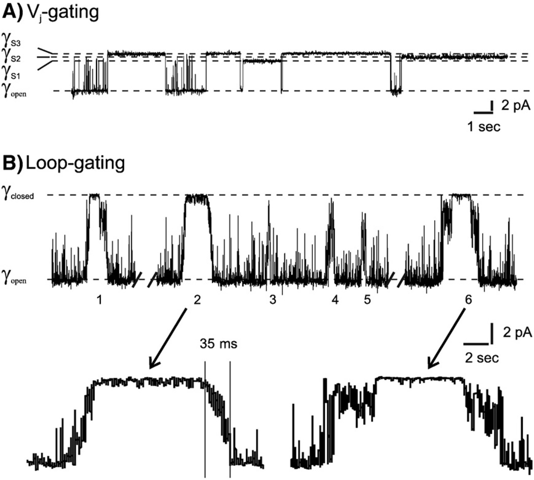
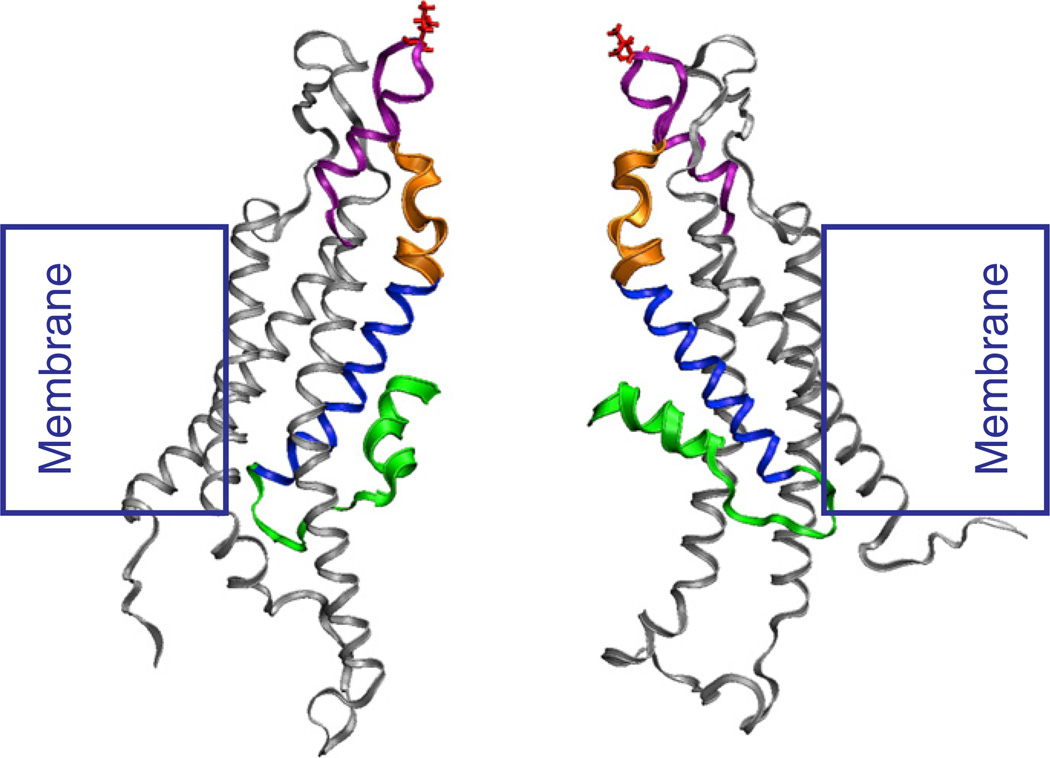
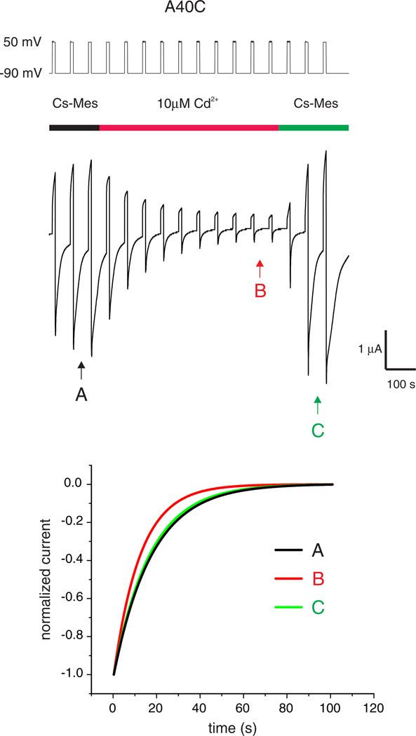
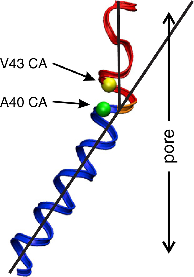
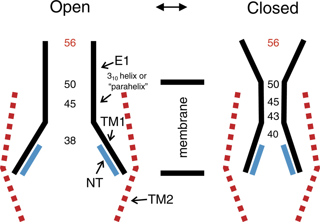
Channels formed by connexins display two distinct types of voltage-dependent gating, termed V(j)- or fast-gating and loop- or slow-gating. Recent studies, using metal bridge formation and chemical cross-linking have identified a region within the channel pore that contributes to the formation of the loop-gate permeability barrier. The conformational changes are remarkably large, reducing the channel pore diameter from 15 to 20Å to less than 4Å. Surprisingly, the largest conformational change occurs in the most stable region of the channel pore, the 3(10) or parahelix formed by amino acids in the 42-51 segment. The data provide a set of positional constraints that can be used to model the structure of the loop-gate closed state. Less is known about the conformation of the V(j)-gate closed state. There appear to be two different mechanisms; one in which conformational changes in channel structure are linked to a voltage sensor contained in the N-terminus of Cx26 and Cx32 and a second in which the C-terminus of Cx43 and Cx40 may act either as a gating particle to block the channel pore or alternatively to stabilize the closed state. The later mechanism utilizes the same domains as implicated in effecting pH gating of Cx43 channels. It is unclear if the two V(j)-gating mechanisms are related or if they represent different gating mechanisms that operate separately in different subsets of connexin channels. A model of the V(j)-closed state of Cx26 hemichannel that is based on the X-ray structure of Cx26 and electron crystallographic structures of a Cx26 mutation suggests that the permeability barrier for V(j)-gating is formed exclusively by the N-terminus, but recent information suggests that this conformation may not represent a voltage-closed state. Closed state models are considered from a thermodynamic perspective based on information from the 3.5Å Cx26 crystal structure and molecular dynamics (MD) simulations. The applications of computational and experimental methods to define the path of allosteric molecular transitions that link the open and closed states are discussed. This article is part of a Special Issue entitled: The Communicating junctions, composition, structure and characteristics.


Figures

Fig. 1
Fig. 1
Single channel records of an undocked Cx32*Cx43E1 hemichannel in Xenopus oocyte. (A) Outside-out record of a single channel illustrating closures by Vj-gating to three substates at a holding potential of −100 mV. (B) Cell-attached recording of a single wild-type channel illustrating six loop-gating events at a holding potential of −70 mV. Note the slow time course of the transitions indicated for events 2 and 6 in the lower panel of B and that not all transitions necessarily lead to full channel closure, as indicated by the partial closure of events 3–5.
Fig. 2
Fig. 2
Macroscopic recording of N2E Cx32*Cx43E1 hemichannel in Xenopus oocytes. Currents shown in the panel on the right were elicited by voltage steps from 0 mV to 40 through −70 mV in 10 mV increments shown in the voltage traces in the left panel. Current relaxations at inside positive potentials (depolarization) result from Vj-gating while those at inside negative potentials (hyperpolarization) result from loop-gating. The N2E substitution reverses the polarity of Vj-gating of the wild type Cx32*Cx43E1 hemichannel. In the wild type channel, current relaxations are only observed with hyperpolarization (not depicted).
Fig. 3
Fig. 3
Coordination geometries of tetradentate cysteine–Cd2+ metal bridges. (A) Tetrahedral coordination geometry showing the Cd2+—S bond distance of 2.5 Å and S—Cd2 +—S bond angle of 109°. All S atoms are separated 4 Å. (B) Planar octahedral geometry, in which the S—Cd2+—S bond angle is 90° and the distance separating all S atoms is 3.5 Å. Only the cysteine side chains are shown. The positions of 2 water molecules that participate in the coordination of Cd2+ (white atom) are shown. Sulfur atoms are yellow spheres, carbon atoms are blue spheres, oxygen atoms are red spheres, nitrogen atoms are blue spheres.
Fig. 4
Fig. 4
State diagrams illustrating the effect of Cd2+ coordination on a voltage dependent two state model. (A). Cd2+ coordination to a binding site created only when the channel adopts the closed conformation. Binding of Cd2+ to the site drives the equilibrium to the right, locking the channel in the closed Cd2+ bound state designated at C*. (B) Cd2+ coordination to a binding site created when the channel adopts the open conformation. Binding of Cd2+ to the site drives the equilibrium to the left, locking the channel in the open Cd2+ bound state designated as O*. The rate constants of voltage dependent transitions between the open O and closed state C are represented as K1 (v) and K−1(v). The voltage dependence of Cd2+ block [91] is represented by the term [Cd2+](v). The potential voltage dependence of “unblock” [92] representing the effect of voltage on the stability of bound Cd2+ is represented by the terms Kon(v) and Koff(v).
Fig. 5
Fig. 5
Side view showing two opposite subunits of a “snapshot” the Cx26 hemichannel following equilibration by MD simulations [11]. The N-terminus is depicted by the green ribbons, with residues 1–11 shown as thick ribbons. The first transmembrane domain, TM1 is depicted by the thin blue ribbons. The 42–51 segment also termed the parahelix, which contains the 310 helix described by Maeda et al. [10] is depicted by thick orange ribbons. This segment contributes to the formation of the loop-gate permeability barrier in Cx32*43E1 and Cx50 hemichannels. The first extracellular loop (E1) is depicted by the thin purple ribbons. It comprises residues 52–71. The position of the 56th residue, which demarcates the extracellular entrance to the channel pore is colored red.
Fig. 6
Fig. 6
Cd2+ lock of Cx32*43E1 A40C hemichannels is reversed by removal of Cd2+ by wash with bath solution. Current traces elicited by voltage steps from −90 to 50 mV are shown. The time of Cd2+ application is indicated by the red line. Wash with bath solution containing 100 mM cesium methanesulfonate (CsMes), 1.8 mM CaCl2 and 10 mM HEPES pH 7.6 reversed Cd2+ lock. The increase in current at the end of the trace most likely results from the incorporation of new channels into the oocyte plasma membrane during the course of the experiment. Kinetics of channel closure before, during and after Cd2+ application (at A, B, and C respectively) are compared in the lower panel. The time constants of loop-gate closure are faster in the presence of Cd2+ as expected if Cd2+ stabilizes the loop-gate closed state (see text).
Fig. 7
Fig. 7
Structure of the TM1/E1 bend angle in a representative Cx26 subunit following equilibration with MD simulation [11]. The positions of the Cα atoms of V43 and A40 are represented by the yellow and green spheres respectively. TM1 is depicted by the blue helix. The segment depicted in red corresponds to residues 41–50, and forms the major part of the loop-gate permeability barrier. The structure shown corresponds to the open channel conformation. In the closed conformation of Cx32*43E1, the geometry of the 41–50 segment changes such that the 43rd and 50th loci move into the channel pore. A40C in 43E1 forms a “lower affinity” Cd2+ binding site, which can be accomplished by straightening the bend angle. The pore diameter formed by the 41–50 segment decreases from ~15–20 to 4 Å in the loop-gate closed state.
Fig. 8
Fig. 8
Schematic representation of the open and loop-gate closed states of a connexin hemichannel, based on studies of conformational changes in the Cx32*Cx43E1 hemichannel. Residues depicted to line the pore of the open channel were determined by substituted cysteine accessibility method using MTSEA-biotin-X as the thiol modifying reagent. The pore diameter in the region of residues depicted in black in the closed model are inferred from the ability of cysteine substitutions at these loci to form metal bridges and/or to be cross-linked by bBBr. The 56th residue is depicted in red to denote that its position does not change substantially with channel closure in either Vj- or loop-gating.

References

    1. Laird DW. Life cycle of connexins in health and disease. Biochem. J. 2006;394:527–543. - PMC - PubMed
    1. Mese G, Valiunas V, Brink PR, White TW. Connexin 26 deafness associated mutations show altered permeability to large cationic molecules. Am. J. Physiol. Cell Physiol. 2008;295:C966–C974. - PMC - PubMed
    1. Bennett MVL, Contreras JE, Bukauskas FF, Sáez JC. New roles for astrocytes: Gap junction hemichannels have something to communicate. Trends Neurosci. 2003;26:610–617. - PMC - PubMed
    1. Saez JC, Retamal MA, Basilio D, Bukauskas FF, Bennett MV. Connexin-based gap junction hemichannels: gating mechanisms. Biochim. Biophys. Acta. 2005;1711:215–224. - PMC - PubMed
    1. Palacios-Prado N, Sonntag S, Skeberdis VA, Willecke K, Bukauskas FF. Gating, permselectivity and pH-dependent modulation of channels formed by connexin 57, a major connexin of horizontal cells in the mouse retina. J. Physiol. 2009;587:3251–3269. - PMC - PubMed

Publication types

MeSH terms

LinkOut - more resources