. 2011 Nov 18;414(1):145-62.
doi: 10.1016/j.jmb.2011.09.023. Epub 2011 Sep 28.

Conservation of lipid functions in cytochrome bc complexes

Affiliations

Conservation of lipid functions in cytochrome bc complexes

S Saif Hasan et al. J Mol Biol. .

Abstract

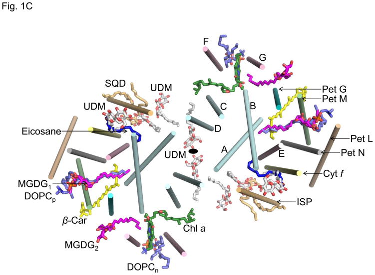
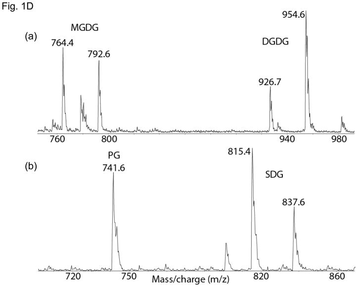
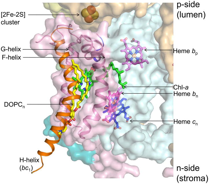
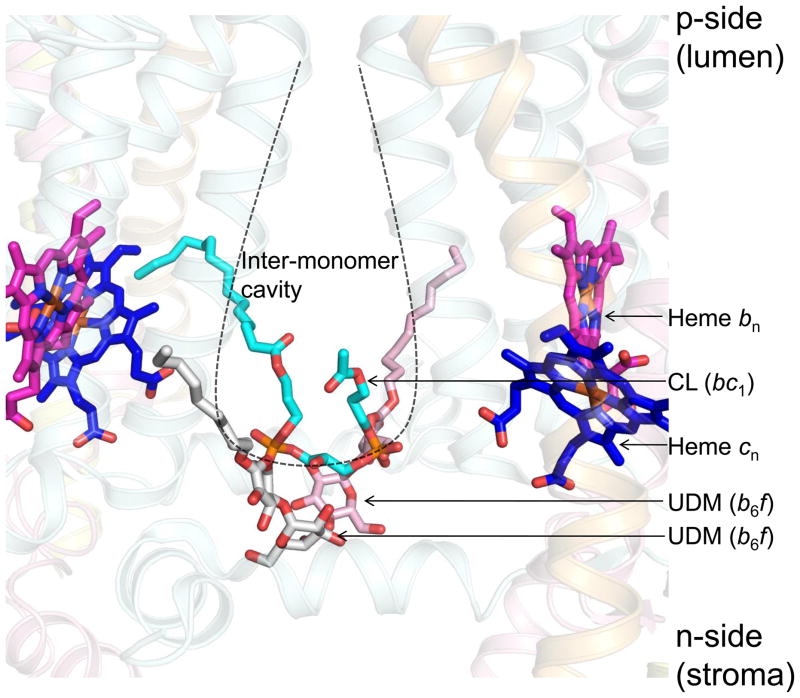
Lipid binding sites and properties are compared in two sub-families of hetero-oligomeric membrane protein complexes known to have similar functions in order to gain further understanding of the role of lipid in the function, dynamics, and assembly of these complexes. Using the crystal structure information for both complexes, we compared the lipid binding properties of the cytochrome b(6)f and bc(1) complexes that function in photosynthetic and respiratory membrane energy transduction. Comparison of lipid and detergent binding sites in the b(6)f complex with those in bc(1) shows significant conservation of lipid positions. Seven lipid binding sites in the cyanobacterial b(6)f complex overlap three natural sites in the Chlamydomonas reinhardtii algal complex and four sites in the yeast mitochondrial bc(1) complex. The specific identity of lipids is different in b(6)f and bc(1) complexes: b(6)f contains sulfoquinovosyldiacylglycerol, phosphatidylglycerol, phosphatidylcholine, monogalactosyldiacylglycerol, and digalactosyldiacylglycerol, whereas cardiolipin, phosphatidylethanolamine, and phosphatidic acid are present in the yeast bc(1) complex. The lipidic chlorophyll a and β-carotene (β-car) in cyanobacterial b(6)f, as well as eicosane in C. reinhardtii, are unique to the b(6)f complex. Inferences of lipid binding sites and functions were supported by sequence, interatomic distance, and B-factor information on interacting lipid groups and coordinating amino acid residues. The lipid functions inferred in the b(6)f complex are as follows: (i) substitution of a transmembrane helix by a lipid and chlorin ring, (ii) lipid and β-car connection of peripheral and core domains, (iii) stabilization of the iron-sulfur protein transmembrane helix, (iv) n-side charge and polarity compensation, and (v) β-car-mediated super-complex with the photosystem I complex.


Figures

Fig 1
Fig 1. Subunit organization and lipid binding sites in the cytochrome b6f complex
(A) Dimeric cytochrome b6f complex from Mastigocladus laminosus (PDB ID 2E74), showing the positions of the eight protein subunits. Side view, in plane of membrane. Color code: Cytochrome f/Pet A (yellow), cytochrome b6/Pet B (cyan), Rieske Fe2S2 protein/Pet C (orange), subunit IV/Pet D (pink), Pet G (teal), Pet L (light brown), Pet M (green) and Pet N (gray). (B) Side view of M. laminosus cyt b6f complex showing lipids, detergents and pigments. (C) Top view along the membrane normal of M. laminosus cytochrome b6f complex (PDB ID 2E74) showing 26 trans-membrane helices and the 2-fold symmetry axis between the monomers. Lipids (MGDG1 and MGDG2, magenta and red) and a pigment (eicosane, blue) from the C. reinhardtii b6f complex were superimposed on the M. laminosus b6f structure by combining PDB ID 2E74 and PDB ID 1Q90. The TMH of cytochrome b6 (A–D) and subunit IV (E–G), Rieske Iron-Sulfur protein, cytochrome f, and the peripheral Pet subunits are shown as cylinders. n, p, electrochemically negative and positive sides of the complex. (D) Neutral and anionic lipids in spinach cyt b6f complex. Major lipids of spinach b6f complex detected by liquid chromatography with mass spectrometry (89). (a). Positive ion mass spectrum of ammoniated neutral lipids (M+NH4)+ after a reverse-phase separation of a chloroform extract of cytochrome b6f complex. The 764.4 and 792.6 Da species are assigned as the (M+NH4)+ ions of monogalactosyldiacylglycerol (MGDG) with 16:3, 18:3 and 18:3, 18:3 fatty acids respectively (C43H70O10 and C45H74O10 calculated mono-isotopic masses 746.49 & 774.53 Da, for the neutral species). The 926.7 and 954.6 Da species are assigned as the (M+NH4)+ ions of digalactosyldiacylglycerol (DGDG) with 16:3, 18:3 and 18:3, 18:3 fatty acids respectively (C49H80O15 and C51H84O15 calculated mono-isotopic masses 908.55 & 936.58 Da, for the neutral species). (b). The negative-ion mass spectrum (m/z 700 – 870) of the same sample is shown. The 741.6 Da species is assigned as the (M-H+) ion of phosphatidylglycerol (PG) with 16:1 and 18:3 fatty acids (C40H71O10P1; calculated mono-isotopic mass 742.48 Da, for the neutral species). The ions at 815.4 and 837.6 Da are assigned as (M-H+) ions of sulfoquinovosyldiacylglycerol (SDG) with either 16:0, 18:3 or 18:3, 18:3 fatty acids respectively (C43H78O12S1 and C45H76O12S1 calculated mono-isotopic masses 816.51 & 838.49 Da, for the neutral species).
Fig. 2
Fig. 2. Substitution of a TMH in bc1 by a lipid and lipid-like chlorophyll a in the b6f complex
C-terminal trans-membrane 8th helix (H-helix, orange) of cyt b subunit of yeast bc1 complex (PDB ID 3CX5) interacts with the F- and G-helices. In superimposed b6f (PDB ID 2E74) and bc1 (PDB ID 3CX5) complexes, a DOPC lipid (yellow acyl tails, orange and red head group), together with the Chl a (green), protrudes from the space between the F- and G- helices (pink) and fills the niche occupied by the H TMH in the bc1 complex. Protein chains of bc1 subunits not shown.
Fig. 3
Fig. 3. Lipid-mediated connection of peripheral monotopic Pet G, L, M, and N subunits and the polytopic core of the b6f complex
The natural galactolipid MGDG1 (green) fills the interstitial space between small Pet G, L, M, and N subunits of the C. reinhardtii cyt b6f complex (PDB ID 1Q90). The artificial lipid DOPCp (stick format, orange) occupies one of the niches between the small Pet subunits in the cyanobacterial cyt b6f complex (PDB ID 2E74). Neighboring β – carotene shown in yellow. A second galactolipid, MGDG2, is found inserted between the TMH of Pet G and the G-helix of sub IV of the algal cyt b6f.
Fig 4
Fig 4. Sub-domain structure of cyt b6f
The cyt b6f monomer (PDB ID 2E74) is organized into two distinct sub-domains- (i) a polytopic sub-domain (shown in pink) that consists of cyt b6 and subIV and constitutes the central portion of the dimer, and (ii) a single TMH sub-domain (shown in blue) that is composed of Pet L, M, N cyt f and ISP. This sub-domain is more peripheral in position. The interface between the two sub-domains is formed by the β–carotene molecule (yellow) and Pet G (teal). View of the cyt b6f complex along the normal to the membrane (2-fold symmetry axis is shown at the inter-monomer interface).
Fig. 5
Fig. 5. Acyl chain stabilization of the trans-membrane helix (TMH) of the Rieske iron-sulfur protein (ISP)
The acyl tails of a phosphatidic acid, PA (yellow, orange) of cyt bc1 is wrapped around the ISP TMH. A UDM (white and red) molecule is located in a similar position in the cyanobacterial b6f complex, while the niche is occupied by eicosane (green) in the algal b6f complex. The cyanobacterial (PDB ID 2E74) and algal (PDB ID 1Q90) b6f complex are superposed with the the yeast bc1 (PDB ID 3CX5).
Fig. 6
Fig. 6. n-side charge compensation
Conserved position for binding acidic lipid(s) (46) on the n-side of cyanobacterial b6f (PDB ID 2E74) and bc1 complexes (PDB ID 3CX5). (A) Three lipid binding sites (1 sulfolipid and 2 UDM molecules) are observed on the n-side of the inter-monomer cavity, close to the lipid-aqueous interface, in each monomer of the cyanobacterial b6f complex (M. laminosus PDB ID 2E74). The head-group of the sulfolipid (color, wheat) is within an interaction distance (3.5 Å) of 1 UDM molecule (white and red), which interacts with another UDM molecule (pink and red, separation 3.8 Å). Disordered acyl chains of sulfolipid and detergent molecules can define a pathway for plastoquinone/plastoquinol exchange between the inter-monomer cavity and bilayer (2Fo-Fc electron density map contoured at σ=1.0 (~0.1 electrons/Å3). The acidic sulfolipid molecule of cyt b6f interacts with Lys275 (cyt f) and Arg16 and Asn20 (ISP). (B) The sulfolipid (wheat, transparent) is located close to the acidic PA (green) and detergent UDM (teal) of the bc1 complex suggesting an important role for this lipid binding niche.
Fig 7
Fig 7. Super-complex formation; bridge to photosystem I
Cardiolipin of cyt bc1 (green and red) is involved in stabilizing super-complexes with cyt c oxidase (55). Superposition of the bc1 complex (PDB ID 3CX5) with the cyanobacterial b6f complex (PDB ID 2E74) results in close spatial location of cardiolipin (acyl chains, green; orange and red are head group phosphorous and oxygen atoms respectively) and β–carotene (yellow), which has been suggested to mediate a cyt b6f-PSI supercomplex (5).
Fig 8
Fig 8. n-side H+ antenna function
An acidic cardiolipin molecule (cyan), shared between the two monomers of the bc1 complex (PDB ID 3CX5). Two detergent molecules (white/red and pink/red, one from each monomer) are found in the same position in the cyanobacterial cyt b6f complex (M. laminosus, PDB ID 2E74) when the structures are superposed and may mark the position of a lipid molecule.

References

    1. Kurisu G, Zhang H, Smith JL, Cramer WA. Structure of the cytochrome b6f complex of oxygenic photosynthesis: tuning the cavity. Science (New York, NY. 2003;302:1009–1014. - PubMed
    1. Yan J, Kurisu G, Cramer WA. Structure of the cytochrome b6f complex: Binding site and intraprotein transfer of the quinone analogue inhibitor 2,5-dibromo-3-methyl-6-isopropyl-p-benzoquinone. Proc Nat Acad Sci USA. 2006;103:67–74. - PMC - PubMed
    1. Yamashita E, Zhang H, Cramer WA. Structure of the cytochrome b6f complex: quinone analogue inhibitors as ligands of heme cn. J Mol Biol. 2007;370:39–52. - PMC - PubMed
    1. Baniulis D, Yamashita E, Whitelegge JP, Zatsman AI, Hendrich MP, Hasan SS, Ryan CM, Cramer WA. Structure-Function, Stability, and Chemical Modification of the Cyanobacterial Cytochrome b6f Complex from Nostoc sp. PCC 7120. J Biol Chem. 2009;284:9861–9869. - PMC - PubMed
    1. Stroebel D, Choquet Y, Popot JL, Picot D. An atypical heam in the cytochrome b6f complex. Nature. 2003;426:413–418. - PubMed

Publication types

MeSH terms

LinkOut - more resources