. 2011 Nov 4;413(4):790-803.
doi: 10.1016/j.jmb.2011.09.024. Epub 2011 Sep 28.

The NLRP12 pyrin domain: structure, dynamics, and functional insights

Affiliations

The NLRP12 pyrin domain: structure, dynamics, and functional insights

Anderson S Pinheiro et al. J Mol Biol. .

Abstract

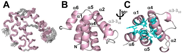
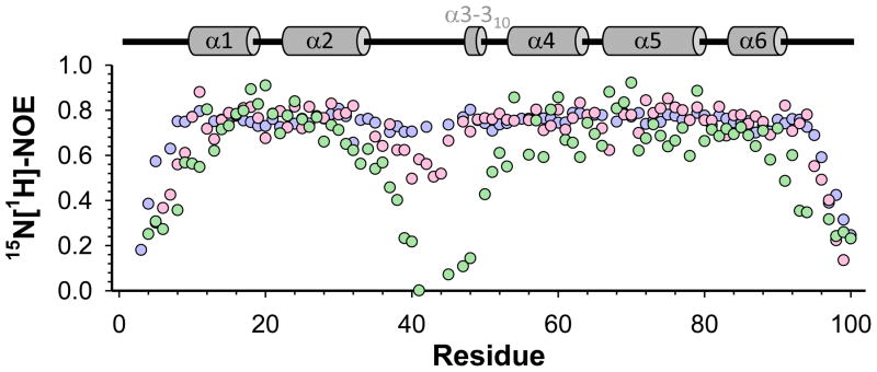
The initial line of defense against infection is sustained by the innate immune system. Together, membrane-bound Toll-like receptors and cytosolic nucleotide-binding domain and leucine-rich repeat-containing receptors (NLR) play key roles in the innate immune response by detecting bacterial and viral invaders as well as endogenous stress signals. NLRs are multi-domain proteins with varying N-terminal effector domains that are responsible for regulating downstream signaling events. Here, we report the structure and dynamics of the N-terminal pyrin domain of NLRP12 (NLRP12 PYD) determined using NMR spectroscopy. NLRP12 is a non-inflammasome NLR that has been implicated in the regulation of Toll-like receptor-dependent nuclear factor-κB activation. NLRP12 PYD adopts a typical six-helical bundle death domain fold. By direct comparison with other PYD structures, we identified hydrophobic residues that are essential for the stable fold of the NLRP PYD family. In addition, we report the first in vitro confirmed non-homotypic PYD interaction between NLRP12 PYD and the pro-apoptotic protein Fas-associated factor 1 (FAF-1), which links the innate immune system to apoptotic signaling. Interestingly, all residues that participate in this protein:protein interaction are confined to the α2-α3 surface, a region of NLRP12 PYD that differs most between currently reported NLRP PYD structures. Finally, we experimentally highlight a significant role for tryptophan 45 in the interaction between NLRP12 PYD and the FAF-1 UBA domain.


Figures

Figure 1
Figure 1
NMR structure of NLRP12 PYD. (A) Ensemble of the 20 lowest-energy structures calculated for NLRP12 PYD superimposed on the backbone atoms of residues 10–91 (PDBID 2L6A). The 6 helices, characteristic of the death domain fold, are highlighted in light pink, while loops are highlighted in grey. (B) Ribbon representation of the lowest-energy conformer of NLRP12 PYD in an orientation identical to that shown in A. The N- and C-termini, as well as the 6 helices are labeled. (C) Top view of NLRP12 PYD (rotated by 90° about the x axis relative to A and B). Residues forming the central hydrophobic core are shown as cyan sticks.
Figure 2
Figure 2
Comparison of fast time scale backbone dynamics among the PYDs of NLRP12, NLRP7 and NLRP1. Comparison of 15N[1H]-NOE (hetNOE) values measured for the PYDs of NLRP12 (light pink), NLRP7 (light blue) and NLRP1 (light green). Experimentally derived secondary structure elements of NLRP12 PYD are depicted by grey cylinders above the figure. A hetNOE of ~0.8 is typical for well-formed, stable secondary structural elements. While the α2-α3 loop and helix α3 in NLRP1 PYD show a substantial increase in fast time scale backbone dynamics, these motions are missing in NLRP7 PYD. The α2-α3 loop and helix α3 in NLRP12 PYD show increased flexibility when compared to NLRP7 PYD.
Figure 3
Figure 3
Sequence alignment of human NLRP PYDs. Residues contributing to the hydrophobic core of NLRP12 PYD are conserved among the entire NLRP family and are highlighted by grey boxes. Experimentally derived secondary structure elements of NLRP12 PYD are depicted by grey cylinders on top of the figure.
Figure 4
Figure 4
Structural comparison of NLRP PYDs. (A) NLRP12 PYD (light pink) overlaid with the PYDs of NLRP7 (light blue; RMSD 2.4 Å) and NLRP1 (light green; RMSD 3.1 Å). The pair wise RMSD between NLRP12 PYD and all other PYDs was calculated by superposition of helices α1-α6. The largest structural difference is localized to the α2-α3 loop as well as helix α3, which is illustrated in the current orientation. (B) Hydrophobic residues in the α2-α3 loop as well as helix α3 are highlighted as dark blue sticks and labeled. A six-residue hydrophobic cluster stabilizes the α2-α3 loop as well as helix α3 in NLRP7 PYD. The corresponding cluster in NLRP12 PYD consists only of 2 hydrophobic residues. NLRP1 PYD lacks all 6 hydrophobic residues. (C) Residues Gly33 and Trp45 in NLRP12 PYD, as well as Trp30 and Trp43 in NLRP7 PYD, are highlighted as dark red sticks and labeled. While Trp43 in NLRP7 PYD is buried and forms stacking interactions with Trp30, the corresponding Trp45 in NLRP12 PYD is surface exposed.
Figure 5
Figure 5
Electrostatics surface potential of NLRP12 PYD and other PYDs with known structures. (A) Top row: ribbon representation of the PYDs of NLRP12, and the ones of the adaptor protein ASC and its inhibitor, ASC2, facing the α2-α3 surface. Bottom row: electrostatic surface representation of the PYDs of NLRP12, ASC and ASC2. Positive surface charge is colored blue; negative surface charge is colored red; and neutral surface, white. (B) Corresponding electrostatic surface representation of the PYDs of NLRP12, NLRP7 and NLRP1.
Figure 6
Figure 6
Mapping the interaction of NLRP12 PYD with FAF-11–57. (A) Chemical shift perturbations (CSPs) calculated for NLRP12 PYD upon titration of FAF-11–57 at a molar ratio of 1:10 (10 mM Na-phosphate buffer pH 7.0, 100 mM NaCl, 0.5 mM TCEP). The color scheme denotes the intensity of the shifts observed in the titration experiment. CSP values higher than two standard deviations from the mean are colored light blue; three standard deviations, marine; and four standard deviations, purple. Experimentally derived secondary structure elements of NLRP12 PYD are depicted by grey cylinders on top of the figure. (B) NLRP12 PYD structure displaying the residues that show the highest CSP values upon titration with FAF-11–57. Side chains are depicted in stick model, labeled and colored according to A. (C) Surface representation of the NLRP12 PYD structure in the same orientation as B, displaying the residues that show the highest CSP values. These residues are clustered on the α2-α3 surface, which has been previously implicated in homotypic interactions of PYDs.
Figure 7
Figure 7
Mapping the interaction of FAF-11–57 with NLRP12 PYD. (A) Chemical shift intensity differences for unbound FAF-11–57 and NLRP12 PYD bound FAF-11–57 (1:10 ratio; 10 mM Na-phosphate buffer pH 7.0, 100 mM NaCl, 0.5 mM TCEP; black bars). Cyan denotes values of I/I0 2× higher than the standard deviation (SD) and blue I/I0 values 3× higher than SD. The color scheme denotes the intensity of the shifts observed in the titration experiment. Experimentally derived secondary structure elements of FAF-11–57 are depicted by grey cylinders above the figure. (B) FAF-15–47 structure (PDBid: 3E21) displaying the residues that show the largest intensity changes upon titration with NLRP12 PYD. Side chains are depicted in stick model, labeled and colored according to A. (C) Surface representation of the FAF-11–57 structure, with residues that show the highest I/I0 differences colored according to A.

References

    1. Fritz JH, Ferrero RL, Philpott DJ, Girardin SE. Nod-like proteins in immunity, inflammation and disease. Nat Immunol. 2006;7:1250–7. - PubMed
    1. Kanneganti TD, Lamkanfi M, Nunez G. Intracellular NOD-like receptors in host defense and disease. Immunity. 2007;27:549–59. - PubMed
    1. Meylan E, Tschopp J, Karin M. Intracellular pattern recognition receptors in the host response. Nature. 2006;442:39–44. - PubMed
    1. Martinon F, Tschopp J. NLRs join TLRs as innate sensors of pathogens. Trends Immunol. 2005;26:447–54. - PubMed
    1. Faustin B, Reed JC. Sunburned skin activates inflammasomes. Trends Cell Biol. 2008;18:4–8. - PubMed

Publication types

MeSH terms

Substances

Associated data