Review
. 2011 Nov;21(11):656-63.
doi: 10.1016/j.tcb.2011.08.008. Epub 2011 Oct 4.

K11-linked ubiquitin chains as novel regulators of cell division

Affiliations
Review

K11-linked ubiquitin chains as novel regulators of cell division

Katherine E Wickliffe et al. Trends Cell Biol. 2011 Nov.

Abstract

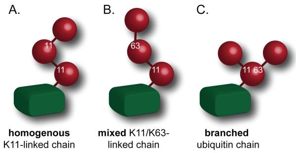
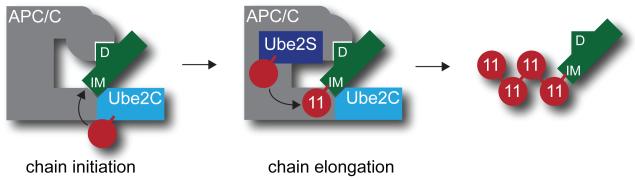
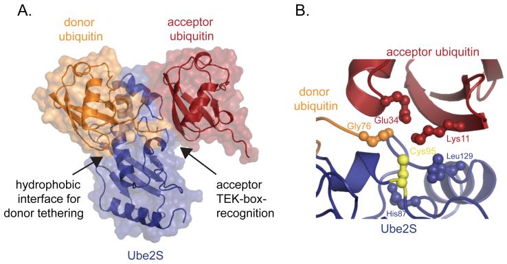
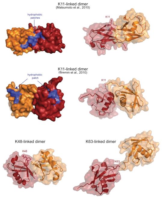
Modification of proteins with ubiquitin chains is an essential regulatory event in cell cycle control. Differences in the connectivity of ubiquitin chains are believed to result in distinct functional consequences for the modified proteins. Among eight possible homogenous chain types, canonical Lys48-linked ubiquitin chains have long been recognized to drive the proteasomal degradation of cell cycle regulators, and Lys48 is the only essential lysine residue of ubiquitin in yeast. It thus came as a surprise that in higher eukaryotes atypical K11-linked ubiquitin chains regulate the substrates of the anaphase-promoting complex and control progression through mitosis. We discuss recent findings that shed light on the assembly and function of K11-linked chains during cell division.


Figures

Figure 1
Figure 1. K11-linkages are found in chains of distinct topologies
A. In homogenous K11-linked chains, all ubiquitin molecules are connected through K11-linkages. B. In mixed chains, K11- and other linkages are found, yet only one amino-group is modified per ubiquitin molecule. C. In branched ubiquitin chains, a single ubiquitin is connected to at least two other ubiquitin molecules.
Figure 2
Figure 2. Mechanism of K11-linked ubiquitin chain formation by the APC/C
Substrates (green) are bound by the APC/C through degrons referred to as D-boxes (D) or KEN-boxes (not shown). Substrates also contain chain initiation motifs (IM) that are recognized by Ube2C, APC/C, or both, and that promote chain initiation by the E2 Ube2C. Following initiation, chains are extended by the K11-specific E2 Ube2S. Ube2C and Ube2S do not compete for binding to the APC/C.
Figure 3
Figure 3. K11-linkage formation occurs through substrate-assisted catalysis
A. Structural model of the ternary complex between Ube2S (blue), donor ubiquitin (orange), and acceptor ubiquitin (red), based on coordinates reported in [45]. The donor ubiquitin is tethered to the E2 by its thioester bond (not shown) and a non-covalent interaction around helix α2 (arrow). The acceptor ubiquitin is recognized through an electrostatic interaction that involves the TEK-box of ubiquitin (arrow). B. The catalytic center of K11-inkage formation consists of residues of the E2 Ube2S (blue) and the substrate acceptor ubiquitin (red). Ube2S contributes the active site cysteine (yellow), Leu129, and His87. The acceptor ubiquitin contributes Glu34 that helps deprotonate Lys11 and orients it towards the active site of Ube2S. The donor ubiquitin is shown in orange.
Figure 4
Figure 4. K11-linked ubiquitin dimers have a unique structure
Two structures of K11-linked ubiquitin dimers, as well as comparable structures of K48- and K63-linked dimers are displayed (based on coordinates reported in pdb-files 3NOB, 2XEW, 2PEA, 2JF5). The acceptor ubiquitin containing the modified lysine residue is shown in red, the donor ubiquitin is depicted in orange. Both K11-linked ubiquitin dimers show compact structures with the hydrophobic patch of ubiquitin being exposed for potential interactions.

References

    1. Schulman BA, Harper JW. Ubiquitin-like protein activation by E1 enzymes: the apex for downstream signalling pathways. Nat Rev Mol Cell Biol. 2009;10(5):319–31. - PMC - PubMed
    1. Ye Y, Rape M. Building ubiquitin chains: E2 enzymes at work. Nat Rev Mol Cell Biol. 2009;10(11):755–64. - PMC - PubMed
    1. Deshaies RJ, Joazeiro CA. RING domain E3 ubiquitin ligases. Annu Rev Biochem. 2009;78:399–434. - PubMed
    1. Deribe YL, Pawson T, Dikic I. Post-translational modifications in signal integration. Nat Struct Mol Biol. 2010;17(6):666–72. - PubMed
    1. Chau V, et al. A multiubiquitin chain is confined to specific lysine in a targeted short-lived protein. Science. 1989;243(4898):1576–83. - PubMed

Publication types

Substances