

Review
doi: 10.1016/j.semcdb.2011.09.010.
Epub 2011 Sep 29.
Neuronal action on the developing blood vessel pattern
Affiliations
- PMID: 21978864
- PMCID: PMC3230733
- DOI: 10.1016/j.semcdb.2011.09.010
Item in Clipboard
Review
Neuronal action on the developing blood vessel pattern
Semin Cell Dev Biol.
2011 Dec.
Abstract
The nervous system relies on a highly specialized network of blood vessels for development and neuronal survival. Recent evidence suggests that both the central and peripheral nervous systems (CNS and PNS) employ multiple mechanisms to shape the vascular tree to meet its specific metabolic demands, such as promoting nerve-artery alignment in the PNS or the development the blood brain barrier in the CNS. In this article we discuss how the nervous system directly influences blood vessel patterning resulting in neuro-vascular congruence that is maintained throughout development and in the adult.
Published by Elsevier Ltd.
Figures
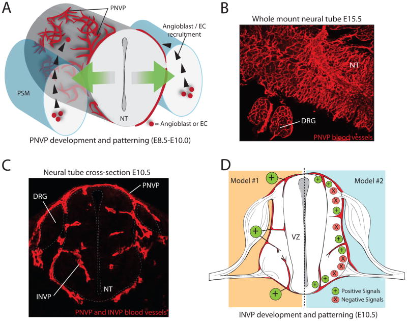
(A) Model of PNVP recruitment. At E8.5, angioblasts and ECs from the PSM respond to positive vessel patterning signals secreted from neural cells (green arrows) by differentiating, proliferating, and migrating to the surface of the neural tube. They surround the neural tube, forming a blood vessel plexus. (B) Immunofluorescence on a whole-mount, E15.5 NT (dorsal view), detecting blood vessels of the PNVP. PNVP vessels form a remodeled network on the surface of the NT and DRG. (C) Immunofluorescence on a cross-section of an E10.5 mouse embryo detecting both PNVP and INVP blood vessels. Blood vessels from the PNVP invade the neural tube at this stage, forming the INVP. (D) Two models for stereotypical vessel invasion during INVP formation. Model #1 depicts positive blood vessel patterning cues (such as matrix-binding VEGF-A) being localized to precise points of blood vessel invasion, whereas Model #2 depicts a balance of positive and negative blood vessel patterning cues to regulate the ingression pattern. Abbreviations: Peri-neural vessel plexus (PNVP), intra-neural vessel plexus (INVP), pre-somitic mesoderm (PSM), dorsal root ganglia (DRG), neural tube (NT), endothelial cell (EC), ventricular zone (VZ).
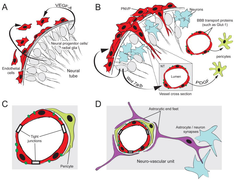
(A) Neural progenitor cells (grey) secrete VEGF-A to recruit ECs to the surface of the neural tube. VEGF-A signaling from the neural tube is also required for subsequent blood vessel ingression into the neural tube (not shown). (B) At the onset of neurogenesis (differentiated neurons in blue), blood vessel sprouts originating from the PNVP invade the neural tube, forming the INVP. Wnt7 signaling is required for proper blood vessel ingression and induction of BBB-specific membrane transport proteins (such as Glut-1). Blood vessels recruit pericytes (light green) via the PDGF-B signaling pathway. (C) Pericyte recruitment is essential for stabilization of the BBB and for maintenance of tight junctions and Glut-1 expression. Pericytes are an important component of the neuro-vascular unit. (D) At the onset of gliogenesis, astrocytes (purple) differentiate and project cellular processes called end -feet to wrap around the CNS blood vessels. Astrocytes also form synaptic complexes with neurons. While some components of the BBB and the neuro-vascular unit develop before astrocytes differentiate, astrocytes are required for stabilization of the BBB and are critical components of the neuro-vascular unit.
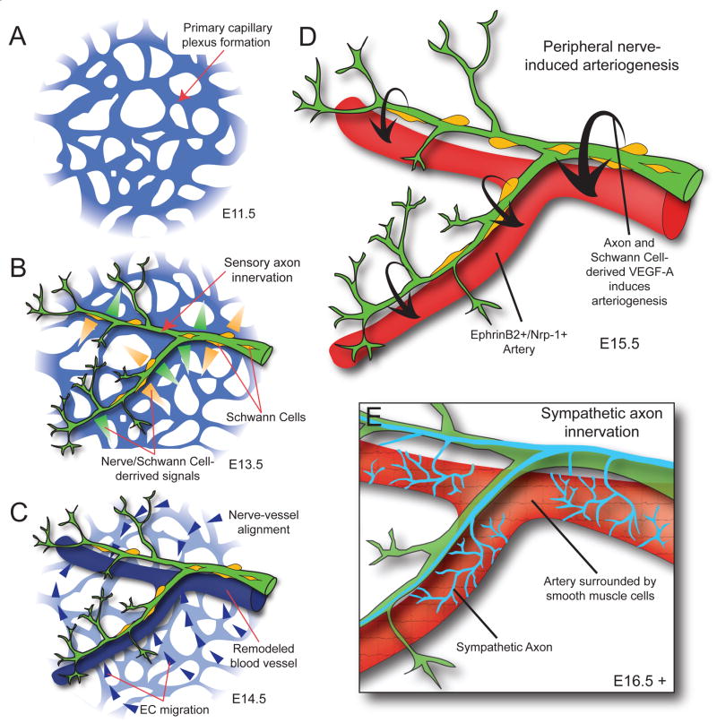
(A) Blood vessels coalesce into a primary capillary plexus and begin to undergo remodeling by E11.5, before peripheral axon innervation in the limb. (B) Sensory axons, and associated Schwann cells, innervate the limb by E13.5 and secrete signals to pattern the primary vascular plexus. (C) In response to nerve and Schwann cell-derived signals, endothelial cells migrate toward and begin to align with peripheral nerves. (D) Blood vessels that have aligned with peripheral nerves undergo arteriogenesis in response to nerve and Schwann cell-derived VEGF-A. Aligned blood vessels upregulate arterial markers such as ephrinB2 and Nrp-1. Up-regulation of Nrp-1 is thought to increase endothelial cell response to VEGF-A signaling. (E) After sensory nerve-induced arteriogenesis is complete, sympathetic axons innervate the limb, utilizing the blood vessels and sensory nerves as a template for migrations. Sympathetic axons invade the arterial smooth muscle layer to regulate local control of vascular tone.
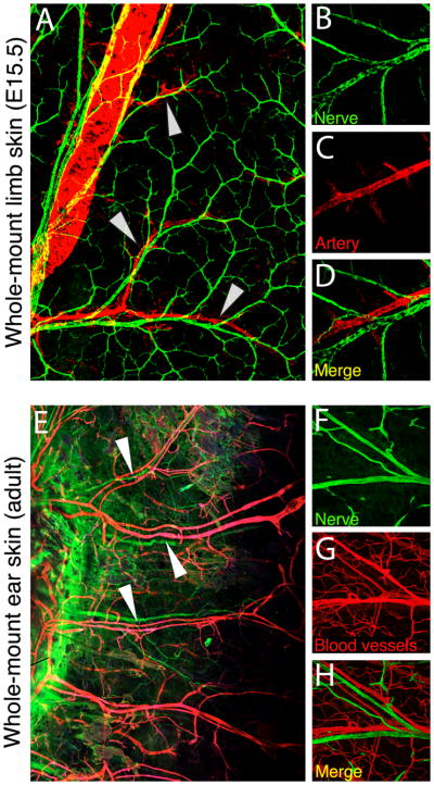
(A–E) Immuno-fluorescence detecting arteries (red) and nerves (green) in whole-mount limb skin preparations from E15.5 mouse embryos (A, 10X and B–D, 20X magnification). Nerves and vessels closely align (arrowheads). (E–H) Immuno-fluorescence detecting blood vessels (red) and nerves (green) in whole-mount ear skin preparations from adult mice (E, 10X and F–H, 20X magnification). Nerve-vessel alignment is maintained in the adult (arrowheads).
References
-
- Stewart PA, Wiley MJ. Structural and histochemical features of the avian blood-brain barrier. J Comp Neurol. 1981;202:157–167. - PubMed
-
- Nakao T, Ishizawa A, Ogawa R. Observations of vascularization in the spinal cord of mouse embryos, with special reference to development of boundary membranes and perivascular spaces. Anat Rec. 1988;221:663–677. - PubMed
-
- Noden DM. Embryonic origins and assembly of blood vessels. Am Rev Respir Dis. 1989;140:1097–1103. - PubMed
-
- Kurz H, Gartner T, Eggli PS, Christ B. First blood vessels in the avian neural tube are formed by a combination of dorsal angioblast immigration and ventral sprouting of endothelial cells. Dev Biol. 1996;173:133–147. - PubMed
-
- Bar T. The vascular system of the cerebral cortex. Adv Anat Embryol Cell Biol. 1980;59(I–VI):1–62. - PubMed
Publication types
MeSH terms
Grants and funding
LinkOut - more resources
Full Text Sources
Other Literature Sources
