. 2012 Apr;19(4):592-604.
doi: 10.1038/cdd.2011.128. Epub 2011 Oct 7.

TRAP1 and the proteasome regulatory particle TBP7/Rpt3 interact in the endoplasmic reticulum and control cellular ubiquitination of specific mitochondrial proteins

Affiliations

TRAP1 and the proteasome regulatory particle TBP7/Rpt3 interact in the endoplasmic reticulum and control cellular ubiquitination of specific mitochondrial proteins

M R Amoroso et al. Cell Death Differ. 2012 Apr.

Abstract

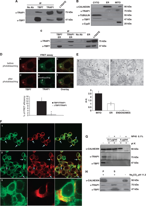
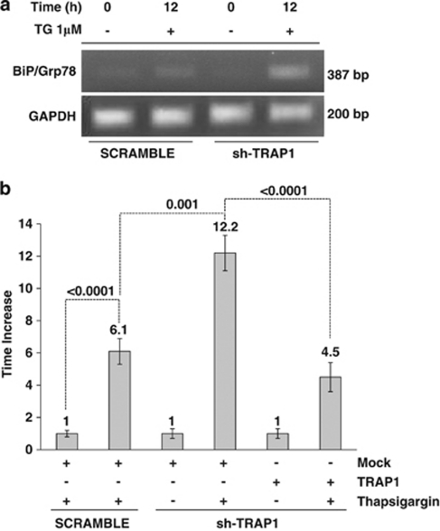
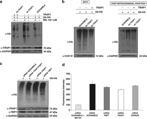
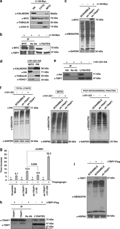
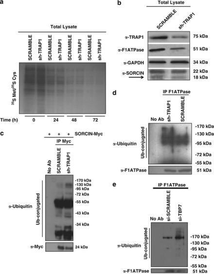
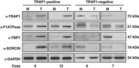
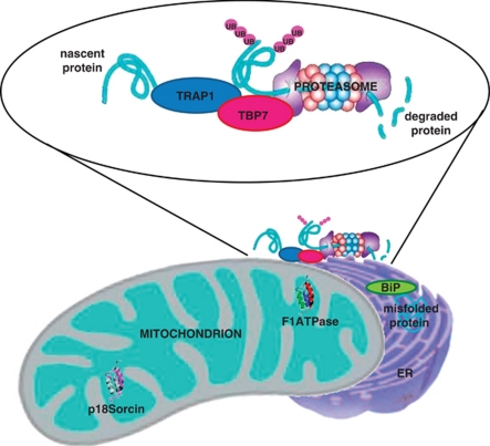
Tumor necrosis factor receptor-associated protein-1 (TRAP1) is a mitochondrial (MITO) antiapoptotic heat-shock protein. The information available on the TRAP1 pathway describes just a few well-characterized functions of this protein in mitochondria. However, our group's use of mass-spectrometric analysis identified TBP7, an AAA-ATPase of the 19S proteasomal subunit, as a putative TRAP1-interacting protein. Surprisingly, TRAP1 and TBP7 colocalize in the endoplasmic reticulum (ER), as demonstrated by biochemical and confocal/electron microscopic analyses, and interact directly, as confirmed by fluorescence resonance energy transfer analysis. This is the first demonstration of TRAP1's presence in this cellular compartment. TRAP1 silencing by short-hairpin RNAs, in cells exposed to thapsigargin-induced ER stress, correlates with upregulation of BiP/Grp78, thus suggesting a role of TRAP1 in the refolding of damaged proteins and in ER stress protection. Consistently, TRAP1 and/or TBP7 interference enhanced stress-induced cell death and increased intracellular protein ubiquitination. These experiments led us to hypothesize an involvement of TRAP1 in protein quality control for mistargeted/misfolded mitochondria-destined proteins, through interaction with the regulatory proteasome protein TBP7. Remarkably, expression of specific MITO proteins decreased upon TRAP1 interference as a consequence of increased ubiquitination. The proposed TRAP1 network has an impact in vivo, as it is conserved in human colorectal cancers, is controlled by ER-localized TRAP1 interacting with TBP7 and provides a novel model of the ER-mitochondria crosstalk.


Figures

Figure 1
Figure 1
TRAP1 and TBP7 interact and colocalize in the ER. (A) Total HCT116 lysates were harvested and immunoprecipitated using anti-TRAP1 and anti-TBP7 antibodies as described under Materials and Methods, separated by SDS-PAGE and immunoblotted using the indicated mouse monoclonal anti-TRAP1 and mouse monoclonal anti-TBP7 antibodies. No Ab, total cellular extracts incubated with A/G plus agarose beads without antibody; IP, immunoprecipitation using the corresponding antibodies. (B) Total HCT116 lysates were fractionated into MITO, CYTO and microsomal (ER) fractions as described under Materials and Methods, separated by SDS-PAGE and immunoblotted using mouse monoclonal anti-TRAP1 and mouse monoclonal anti-TBP7 antibodies. The purity of the fractions was assessed by using mouse monoclonal anti-tubulin, goat polyclonal anti-CypD, rabbit polyclonal anti-calnexin antibodies specific for the single subcellular compartments. (C) TRAP1 and TBP7 co-IP analysis on the microsomal fraction (ER), obtained as described under Materials and Methods. WB of immunoprecipitates was performed by using the indicated antibodies. (D) TRAP1/TBP7 direct interaction. FRET was measured by using the acceptor photo-bleaching technique as described under Materials and Methods. The images show the signal of TBP7 (red) and TRAP1 (green) before (a–c) and after photo-bleaching (d–f). The selected ROI for bleaching was indicated. Energy transfer efficiency was measured in cells transiently co-transfected with TRAP1 and either TBP7 or its mutant form (ΔTBP7-Flag), and is expressed in % as mean of three independent experiments. Error bars: ±S.D.; *P<0.0001. (E) ER Distribution of TRAP1 in HCT116 cells (EM). Cells expressing TRAP-HA vector were fixed and prepared for immuno-EM (see Materials and Methods). Labeling with the anti-HA antibody revealed significant amount of TRAP1 in mitochondria (a, b, arrowheads). In addition, TRAP1 was distributed throughout the elongated membrane profiles (a, arrows) that on the basis of their ultrastructural features (such as attached ribosomes) can be attributed to the rough ER compartment, and detected along the nuclear envelope (b, arrows). The density of immuno-gold labeling (in arbitrary units; average±S.D.) in mitochondria (MITO), ER and endosomes (as a negative control) is reported in the lower histogram. (F) ER TRAP1/TBP7 colocalization (confocal microscopy). Immunofluorescence shows colocalization of TBP7 with TRAP1 and with the ER protein calnexin. In Panel-1, a double immunofluorescent staining is shown for TRAP1 (green) and TBP7 (red). In Panel-2, a double immunofluorescent staining is shown for calnexin (green) and TBP7 (red). In cells expressing the Myc-tagged TRAP1 construct (red) the protein co-distributes to a great extent with endogenous calnexin (green, Panel-3). (G and H) Biochemical characterization of TRAP1/TBP7 ‘topology' in the ER. WB of HCT116 microsomal fractions treated with 0.4 μg/ml or 4 μg/ml proteinase-K (pt K) ±1% NP-40 for 20 min on ice (G) or with 100 mM Na2CO3 (pH 11.3) for 30 min (H) as described under Materials and Methods. Specific proteins were revealed using the indicated antibodies. (H): S, supernatant; P, pellet
Figure 2
Figure 2
BiP mRNA levels in sh-TRAP1 stable clones. (a) Semi-quantitative RT-PCR analysis of BiP/Grp78 mRNA expression in sh-TRAP1 stable clones with respect to scrambled transfectants after 12-h treatment with 1 μM TG. As control, the levels of GAPDH transcript were analyzed. (b) Real-time RT-PCR analysis of BiP/Grp78 mRNA expression in scrambled and sh-TRAP1 HCT116 cells exposed to 1 μM TG for 12 h and in sh-TRAP1 HCT116 cells transfected with TRAP1 cDNA before treatment with TG. The P-values indicate the statistical significance between different BiP/Grp78 levels under the indicated conditions
Figure 3
Figure 3
Ub levels in HCT116 cells. (a) Total cell lysates from sh-TRAP1 and scrambled HCT116 stable clones were transfected with either an HA-tagged Ub vector (Ub-HA) or with TRAP1 expression vectors; treated with 1 μM MG132 for 24 h; harvested 48 h after transfection; and subjected to immunoblot using rabbit polyclonal anti-HA antibodies. The same filter was re-probed using mouse monoclonal anti-GAPDH antibodies for normalization of cell lysates. Three independent experiments were performed, with similar results. (b) Sub-cellular fractionation was obtained from sh-TRAP1 and scrambled HCT116 stable transfectants treated as described in panel a. The extracts from the PM fraction (microsomes+CYTO fraction) and mitochondria (MITO, see Materials and Methods) were separated by SDS-PAGE and immunoblotted using a rabbit polyclonal anti-HA antibody to detect Ub levels. The purity of fractions was verified by using mouse monoclonal anti-COX IV or mouse monoclonal anti-GAPDH antibodies. Three independent experiments were performed, with similar results. (c) HCT116 cells were co-transfected with a Ub-HA vector and an siRNA negative control (scramble), or with siRNAs specific for TRAP1, TBP7, or both (as indicated) and total cell lysates were harvested after 48 h from transfection. Total lysates were subjected to SDS-PAGE and immunoblotted using rabbit polyclonal anti-HA antibodies to detect total Ub levels. The same filter was re-probed using mouse monoclonal anti-GAPDH antibodies for normalization of cell lysates, and using mouse monoclonal anti-TRAP1 and mouse monoclonal anti-TBP7 antibodies. (d) Proteasome activity is not affected by TRAP1 and TBP7 silencing. Total cellular extracts were prepared after 48 h of transfection with specific siRNA for TRAP1, TBP7 or Sorcin, as control, or with an siRNA negative control (scramble), and incubated in the presence of assay buffer and the fluorogenic substrate Suc-LLVY-AMC, as described under Materials and Methods. Samples were analyzed in triplicate using an excitation wavelength of 360 nm and an emission wavelength of 450 nm to detect chymotryptic proteasome activity. The data represent the mean of three independent experiments
Figure 4
Figure 4
The TRAP1/TBP7 interaction in the ER is required for control of protein ubiquitination and ER stress. (a and d) Sub-cellular localization of Δ1–59-Myc/Δ101–221-HA mutants. HCT116 cells were transfected with the Δ1–59-Myc (a) or Δ101–221-HA (d) TRAP1 mutants; sub-fractionated into MITO, CYTO and microsomal (ER) fractions (a), or MITO and PM (cytosol+microsomes) fractions (d), as described under Materials and Methods; separated by SDS-PAGE; and immunoblotted using the indicated antibodies to verify the expression of mutants and the purity of fractions. For details on procedures for generation of the mutants see Materials and Methods. (b, e) Interaction between Δ1–59-Myc/Δ101–221-HA mutants and TBP7. HCT116 cells were transfected with Δ1–59-Myc (b) or Δ101–221-HA (e) TRAP1 mutants, harvested and immunoprecipitated using anti-Myc or anti-HA antibodies as described under Materials and Methods. Immunoprecipitates were separated by SDS-PAGE and immunoblotted using the indicated antibodies. No Ab, total cellular extracts incubated with A/G plus agarose beads without antibody; IP, immunoprecipitation with the corresponding antibodies. Three independent experiments were performed, with similar results. (c) Ubiquitination levels upon transfection of the Δ1–59-Myc TRAP1 deletion mutant. Total lysates from HCT116 scrambled, sh-TRAP1 stable clones and sh-TRAP1 cells transfected with the Δ1–59-Myc TRAP1 mutant were subjected to immunoblot analysis using mouse monoclonal anti-Ub antibodies to detect total ubiquitination levels and with an anti-GAPDH antibody for normalization of cell lysates. Three independent experiments were performed, with similar results. (f) Ubiquitination levels upon transfection of the Δ101–221-HA TRAP1 deletion mutant. HCT116 scramble, sh-TRAP1 and sh-TRAP1 cells transfected with the Δ101–221-HA TRAP1 mutant were sub-fractionated in PM (microsomes+CYTO fraction) and MITO fractions as described under Materials and Methods. Total lysates from the same cells were used as controls (left panel). Protein lysates were subjected to immunoblot analysis using mouse monoclonal anti-Ub antibodies to detect total ubiquitination levels. The purity of fractions was verified using mouse monoclonal anti-GAPDH (left and right panels) and mouse-monoclonal anti-COX IV (middle panel) antibodies. Three independent experiments were performed, with similar results. (g) Real-time RT-PCR analysis of BiP/Grp78 mRNA expression in scrambled and sh-TRAP1 HCT116 cells exposed to 1 μM TG for 12 h (same as in Figure 2b) and in sh-TRAP1 HCT116 cells transfected with the Δ1–59-Myc or Δ101–221-HA TRAP1 mutant, as indicated, before treatment with TG. The P-values indicate the statistical significance between the BiP/Grp78 levels under the indicated conditions. (h) Interaction between TRAP1 and the ΔTBP7-Flag deletion mutant. HCT116 cells were transfected with the ΔTBP7-Flag deletion mutant, harvested and immunoprecipitated using anti-TRAP1 antibodies as described under Materials and Methods. Immunoprecipitates were separated by SDS-PAGE and immunoblotted using the indicated antibodies. No Ab, total cellular extracts incubated with A/G plus agarose beads without antibody; IP, immunoprecipitation with the corresponding antibodies. Three independent experiments were performed, with similar results. The arrow indicates the ΔTBP7-Flag mutant band. (i) Ubiquitination levels upon transfection of the ΔTBP7-Flag deletion mutant. Total lysates from HCT116 scrambled cells transfected with ΔTBP7-Flag mutant were subjected to immunoblot analysis using mouse monoclonal anti-Ub antibodies to detect total ubiquitination levels and with mouse monoclonal anti-HSP60 antibodies for normalization of cell lysates. Three independent experiments were performed, with similar results
Figure 5
Figure 5
Control of intracellular protein stability and ubiquitination pattern of p18 Sorcin and F1ATPase. (a) Pulse–chase analysis of total lysates of scrambled and sh-TRAP1 HCT116 cells. HCT116 cells were incubated in cysteine/methionine-free medium for 1 h followed by incubation in cysteine/methionine-free medium containing 50 μCi/ml 35S-labeled cysteine/methionine (35S Met/35S Cys) for 1 h. After labeling, cells were washed once with culture medium containing 10-fold excess of unlabeled methionine and cysteine (5 mM each) and incubated further in the same medium for the indicated time periods. Cells were collected at the indicated time points and total lysates subjected to SDS-PAGE were analyzed by autoradiography. (b) Total lysates of scrambled and sh-TRAP1 HCT116 cells were subjected to SDS-PAGE and immunoblotted using rabbit polyclonal anti-Sorcin, mouse monoclonal anti-TRAP1, goat polyclonal anti-F1ATPase antibodies. The same filter was re-probed using mouse monoclonal anti-GAPDH antibodies for normalization of cell lysates. The arrow indicates the MITO 18-kDa Sorcin isoform band. (c) Scrambled and sh-TRAP1 HCT116 clones were transfected with an expression vector containing the cDNA of p18 Sorcin fused to a c-Myc epitope at the C-terminus (Sorcin-Myc) and treated with 1 μM MG132 for 24 h before harvesting. Lysates were immunoprecipitated using mouse monoclonal anti-Myc antibodies and analyzed by immunoblot analysis using mouse monoclonal anti-Ub antibodies. The membrane was re-probed using an anti-Myc antibody to control transfection efficiency. (d) Scrambled and sh-TRAP1 HCT116 clones were treated with 1 μM MG132 for 24 h before harvesting, immunoprecipitated with an-anti F1ATPase antibody, subjected to SDS-PAGE and immunoblotted using mouse monoclonal anti-Ub and goat polyclonal anti-F1ATPase antibodies. Three independent experiments were performed with similar results. (e) HCT116 cells were co-transfected with a Ub-HA vector and an siRNA negative control (scramble), or with siRNAs specific for TBP7; treated with 1 μM MG132 for 24 h before harvesting; immunoprecipitated using a goat polyclonal anti-F1ATPase antibody; subjected to SDS-PAGE; and immunoblotted using mouse monoclonal anti-Ub and goat polyclonal anti-F1ATPase antibodies. Three independent experiments were performed, with similar results
Figure 6
Figure 6
TRAP1, TBP7, F1ATPase and Sorcin expression in human CRCs. Total cell lysates from four human CRCs (T) and the respective non-infiltrated peri-tumoral mucosas (M) were separated by SDS-PAGE and immunoblotted using rabbit polyclonal anti-Sorcin, mouse monoclonal anti-TRAP1, mouse monoclonal anti-TBP7 and goat polyclonal anti-F1ATPase. The same filter was re-probed using mouse monoclonal anti-GAPDH antibodies for normalization of cell lysates. For details on case numbers, refer to Supplementary Figure 1
Figure 7
Figure 7
Crosstalk between ER and mitochondria, and MITO protein quality control. TRAP1 forms a supra-molecular complex with TBP7 on the outside of the ER, in a cellular compartment of tight ER–mitochondria contact sites, where proteasomes are also present. This TRAP1/TBP7 complex is involved in the control of protein stability and intracellular protein ubiquitination of mitochondria-destined proteins. These two proteins, each with independent but related functions, help to judge whether a protein can be repaired and reach the final MITO destination or, if damaged, needs to be degraded through the Ub–proteasome system

References

    1. Song HY, Dunbar JD, Zhang YX, Guo D, Donner DB. Identification of a protein with homology to HSP90 that binds the type 1 tumor necrosis factor receptor. J Biol Chem. 1995;270:3574–3581. - PubMed
    1. Felts SJ, Owen BA, Nguyen P, Trepel J, Donner DB, Toft DO. The HSP90-related protein TRAP1 is a mitochondrial protein with distinct functional properties. J Biol Chem. 2000;275:3305–3312. - PubMed
    1. Montesano Gesualdi N, Chirico G, Catanese MT, Pirozzi G, Esposito F. AROS-29 is involved in adaptive response to oxidative stress. Free Radic Res. 2006;40:467–476. - PubMed
    1. Montesano Gesualdi N, Chirico G, Pirozzi G, Costantino E, Landriscina M, Esposito F. Tumor necrosis factor-associated protein 1 (TRAP-1) protects cells from oxidative stress and apoptosis. Stress. 2007;10:342–350. - PubMed
    1. Costantino E, Maddalena F, Calise S, Piscazzi A, Tirino V, Fersini A, et al. TRAP1, a novel mitochondrial chaperone responsible for multi-drug resistance and protection from apoptosis in human colorectal carcinoma cells. Cancer Lett. 2009;279:39–46. - PubMed

Publication types

MeSH terms

Substances