Review
. 2011 Oct 1;25(19):2011-24.
doi: 10.1101/gad.169037.

Mechanisms and pathways of growth failure in primordial dwarfism

Affiliations
Review

Mechanisms and pathways of growth failure in primordial dwarfism

Anna Klingseisen et al. Genes Dev. .

Abstract

The greatest difference between species is size; however, the developmental mechanisms determining organism growth remain poorly understood. Primordial dwarfism is a group of human single-gene disorders with extreme global growth failure (which includes Seckel syndrome, microcephalic osteodysplastic primordial dwarfism I [MOPD] types I and II, and Meier-Gorlin syndrome). Ten genes have now been identified for microcephalic primordial dwarfism, encoding proteins involved in fundamental cellular processes including genome replication (ORC1 [origin recognition complex 1], ORC4, ORC6, CDT1, and CDC6), DNA damage response (ATR [ataxia-telangiectasia and Rad3-related]), mRNA splicing (U4atac), and centrosome function (CEP152, PCNT, and CPAP). Here, we review the cellular and developmental mechanisms underlying the pathogenesis of these conditions and address whether further study of these genes could provide novel insight into the physiological regulation of organism growth.


Figures
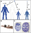
Figure 1.
Figure 1.
(A) Cell size and cell number determine organism size. Conceptually, body size can be altered through reducing the number of cell divisions or reducing cell size. For example, if during an identical period of development, cells divide only five times out of the usual seven rounds of cell division, this will reduce body volume by 75%. Reducing cell volume to a quarter of normal could similarly reduce body size while maintaining cell number constant. (B) In mammals, body size appears to be predominantly determined by cell number. There is a 3000-fold difference in body mass between mice (25 g) and humans (70 kg), while volume of cells from similar tissues remains relatively unchanged (Conlon and Raff 1999). (C) Cell number can be increased in mammals through alterations in proliferation kinetics. For instance, transgenic expression of stabilized β-catenin protein enlarges brain size in mice during embryogenesis. Midcoronal sections through the embryonic day 15.5 (E15.5) cerebral cortex from a control mouse embryo (left) and a mouse with the Δ90β-catenin-GFP transgene expressed in neural precursors, resulting in an enlarged brain with increased cerebral cortical surface area and folds resembling sulci and gyri of higher mammals. Bar, 1 mm. (Image from Chenn and Walsh 2002. Reprinted with permission from AAAS.)
Figure 2.
Figure 2.
Intracellular signaling pathways regulating growth. PI3K/TOR, Hippo, and MAPK pathways regulate growth by modulating protein translation, cell cycle progression, and apoptosis. Schematic of pathways showing key components. Genes highlighted in red are mutated in human genetic syndromes that manifest growth deficiency or overgrowth. (A) Growth hormone acts systemically through its regulation of IGF-1, which activates phosphotidyl-inosine-3 kinase (PI3K) by binding the IGF-1 receptor (IGF-1R). Subsequent activation of downstream kinases results in increased protein translation and ribosome biosynthesis, leading to cellular growth. The pathway is inhibited by the phosphatase PTEN and integrates multiple other signals, such as nutrient/energy levels, through the master kinase target of rapamycin (TOR). TOR activates the ribosomal S6 kinase and facilitates eIF4E activity to promote translation and transcription initiation. (B) The Hippo pathway restricts growth to control organ size and prevent tissue overgrowth and tumorigenesis. The pathway is currently best defined in Drosophila, where the cell polarity protein Crumbs (Crb) and the protocadherins Fat and Dachsous (Ds) activate Hippo kinase, which in turn activates Warts kinase. The signaling cascade negatively regulates the transcriptional coactivator Yorkie (Yki) by retaining Yki in the cytoplasm. This restricts cell proliferation and promotes cell death, as Yki promotes G1 progression over G0 cell cycle exit through transcriptional up-regulation of Cyclin E (CycE) and the E2F transcription factor. Yki also has an anti-apoptotic effect by inducing inhibitor of apoptosis protein (IAP). The core pathway is conserved in mammals: Mst1/2 (Hippo), Lats (Warts), and Yap (Yki). Homologs to Fat and Ds or the target gene bantam have not yet been identified in mammals. (C) The MAPK (ERK) signaling cascade transduces mitogen signals, driving cellular proliferation by promoting G1-to-S-phase progression (Meloche and Pouysségur 2007). Downstream, ERK kinase activates the proproliferative transcription factors Myc and E2F as well as decreases levels of the cyclin-dependent kinase inhibitors p21 and p27. Rather than being entirely discrete signaling pathways, these three signaling pathways (A–C) overlap; for instance, Akt inhibits Hippo activity, while ERK phosphorylates, and thus activates, TOR.
Figure 3.
Figure 3.
Primordial dwarfism is a disorder of extreme global growth failure. Diagram illustrating the proportionate scaling of body structures in primordial dwarfism (A), relative to those of an adult of average stature (B). (C) Reduced head size distinguishes primordial dwarfism from other forms of dwarfism (e.g., achondroplasia). Bar, 25 cm.
Figure 4.
Figure 4.
Cellular pathways implicated in primordial dwarfism. (A) ATR DNA damage response signaling. The ATR kinase is activated in response to ssDNA generated by DNA damage. It phosphorylates downstream targets, including the effector kinase Chk1, to coordinate cell cycle arrest, repair the damage, and/or induce apoptosis. Phosphorylation of Chk1 is required for its accumulation at the centrosome (Niida et al. 2007), where it inhibits Cdc25B phosphatase, preventing Cdk1/Cyclin B activation and the transition from G2 to mitosis (Kramer et al. 2004). MCPH1 and PCNT are required for Chk1 localization at the centrosome, and therefore mutations in these genes also impair ATR-induced G2/M checkpoint signaling. Another primordial dwarfism gene, CEP152, interacts with ATR through binding of CINP, which in turn interacts with ATRIP. Genes mutated in primordial dwarfism/primary microcephaly are highlighted in white text. (B) Licensing of replication origins. During G1, the six-subunit ORC assembles at origins of replication. Once the ORC complex is formed, additional factors such as CDC6 and CDT1 are recruited, permitting reiterative loading of the MCM helicase complex (MCM2–7). After G1/S-phase transition, DNA replication is then initiated by the binding of additional factors and the MCM helicase unwinding DNA at replication forks. (C) mRNA splicing. Splicing of a subpopulation of mRNAs that contain U12-type introns is dependent on the minor spliceosome. The small nuclear ncRNA U4atac is one component of this alternative spliceosome, and mutations in U4atac impair splicing of such introns to a variable degree. U12-dependent introns are found in many genes, including those involved in DNA replication and growth, such as ORC3, MAPKs 1–4, PTEN, and PI3K adaptor protein 1. The exon junction complex (EJC) protein Magoh has also recently been found to be required for splicing of specific genes, including mapk in Drosophila and Lis1 in mice, where it causes microcephaly and growth retardation.
Figure 5.
Figure 5.
Primordial dwarfism genes act in processes regulating cell cycle progression. Schematic of nuclear and centrosome cycles. Centrosome and genomic DNA duplication are coordinately regulated, with both occurring only once per cell cycle. Mutations in preRC complex proteins impair G1/S transition and S-phase progression. They may also increase replication stress through fewer licensed origins being present. ATR also regulates S-phase progression, as ATR-Seckel mice have increased replication stress during embryogenesis. ATR is then required during G2/M transition, signaling via Chk1 at the centrosome, with localization of Chk1 dependent on MCPH1 and PCNT. PCNT and CEP152 are required for nucleation of the mitotic spindle. Additionally, CEP152 and CPAP (CENPJ), as components of the centriole biogenesis machinery, are essential for centriole duplication, which may also impact on mitotic centrosome integrity later in the cell cycle.

References

    1. Alarcon C, Zaromytidou AI, Xi Q, Gao S, Yu J, Fujisawa S, Barlas A, Miller AN, Manova-Todorova K, Macias MJ, et al. 2009. Nuclear CDKs drive Smad transcriptional activation and turnover in BMP and TGF-β pathways. Cell 139: 757–769 - PMC - PubMed
    1. Alderton GK, Joenje H, Varon R, Borglum AD, Jeggo PA, O'Driscoll M 2004. Seckel syndrome exhibits cellular features demonstrating defects in the ATR-signalling pathway. Hum Mol Genet 13: 3127–3138 - PubMed
    1. Alderton GK, Galbiati L, Griffith E, Surinya KH, Neitzel H, Jackson AP, Jeggo PA, O'Driscoll M 2006. Regulation of mitotic entry by microcephalin and its overlap with ATR signalling. Nat Cell Biol 8: 725–733 - PubMed
    1. Al-Dosari MS, Shaheen R, Colak D, Alkuraya FS 2010. Novel CENPJ mutation causes Seckel syndrome. J Med Genet 47: 411–414 - PubMed
    1. Alioto TS 2007. U12DB: a database of orthologous U12-type spliceosomal introns. Nucleic Acids Res 35: D110–D115 doi: 10.1093/nar/gkl796 - PMC - PubMed

Publication types