. 2011 Oct 1;25(19):2031-40.
doi: 10.1101/gad.16872411.

Essential role for DNA-PK-mediated phosphorylation of NR4A nuclear orphan receptors in DNA double-strand break repair

Affiliations

Essential role for DNA-PK-mediated phosphorylation of NR4A nuclear orphan receptors in DNA double-strand break repair

Michal Malewicz et al. Genes Dev. .

Abstract

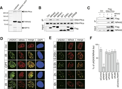
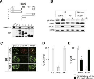
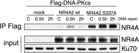
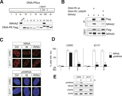
DNA-dependent protein kinase (DNA-PK) is a central regulator of DNA double-strand break (DSB) repair; however, the identity of relevant DNA-PK substrates has remained elusive. NR4A nuclear orphan receptors function as sequence-specific DNA-binding transcription factors that participate in adaptive and stress-related cell responses. We show here that NR4A proteins interact with the DNA-PK catalytic subunit and, upon exposure to DNA damage, translocate to DSB foci by a mechanism requiring the activity of poly(ADP-ribose) polymerase-1 (PARP-1). At DNA repair foci, NR4A is phosphorylated by DNA-PK and promotes DSB repair. Notably, NR4A transcriptional activity is entirely dispensable in this function, and core components of the DNA repair machinery are not transcriptionally regulated by NR4A. Instead, NR4A functions directly at DNA repair sites by a process that requires phosphorylation by DNA-PK. Furthermore, a severe combined immunodeficiency (SCID)-causing mutation in the human gene encoding the DNA-PK catalytic subunit impairs the interaction and phosphorylation of NR4A at DSBs. Thus, NR4As represent an entirely novel component of DNA damage response and are substrates of DNA-PK in the process of DSB repair.


Figures

Figure 1.
Figure 1.
NR4A receptors interact with DNA-PKcs and are recruited to DNA repair foci after DNA damage. (A) Silver staining of material copurifying with affinity-captured tagged NR4A2 (T-NR4A2) or mutant NR4A2 where the last 12 C-terminal amino acids were deleted (T-NR4A2 mAF2). (B) Immunoprecipitation of Flag-tagged nuclear receptors with endogenous DNA-PKcs from nuclear extracts from HEK 293 cells transfected with the indicated expression vectors. (C) Interaction of NR4A2 and Flag-tagged DNA-PKcs in HEK 293 cells transfected with expression vectors for NR4A2 and Flag-DNA-PKcs, as indicated. Cells were either untreated or exposed to IR (3 Gy) as indicated. (D, top panel) Immunofluorescence images of primary MEFs stained against NR4A (red) or γH2AX (green). Antibodies against 53BP1 were used in the bottom panel (green). Cells were untreated (control), exposed to IR (3 Gy), or treated with camptothecin for 30 min (CPT, 10 μm). Cells were fixed and imaged 30 min after IR treatment or camptothecin washout. (E) Immunofluorescence images of normal human fibroblasts (C5RO cells). Cells were exposed to IR (1 Gy), fixed, and stained for NR4A (red) and γH2AX (green) at the indicated time points. (F) Translocation of NR4A to DSB foci requires PARP-1. Quantification of NR4A translocation to DSB foci after 2 Gy of irradiation in U2OS cells transfected with siRNAs or treated with PARP inhibitor (PARPi) as indicated. The graph shows a percentage of γH2AX-positive foci that are also NR4A-positive at 30 min post-irradiation. Error bars indicate SD; n = 3; (*) significant at P < 0.05; (**) significant at P < 0.01.
Figure 2.
Figure 2.
NR4A receptors regulate DSB repair in mammalian cells. (A) Antibodies against either γH2AX or H2AX were used in immunoblots detecting proteins from MEF cell extracts from cells treated as indicated. (B) Knockout NR4A2 MEFs (NR4A2−/−) were infected with GFP or NR4A2 lentiviruses. Cells were treated with camptothecin and used in immunoblotting as in A. (C) MEFs or U2OS cells were exposed to IR (15 Gy), and DNA repair was quantified by the neutral comet assays. Wild-type (wt) cells or NR4A2 knockout (NR4A2−/−) MEFs were infected with control shRNA (shControl) or NR4A1 shRNA (shNR4A1) lentiviruses. Error bars indicate SD; n = 4; (*) significant at P < 0.05 calculated against wild-type shControl sample with the corresponding time of DNA repair. U2OS cells were transfected with siRNAs (siControl; siNR4A1; siNR4A2) as indicated. The diagrams illustrate the percentage of DNA detected in comet tails. Error bars indicate SD; n = 4; (**) significant at P < 0.01 calculated against the siControl sample with the corresponding time of DNA repair. (D) DNA repair foci resolution kinetics at the indicated time points in U2OS cells exposed to IR (1 Gy). U2OS cells were transfected with the same siRNAs as those used in C. Error bars indicate SD; n = 3; (*) significant at P < 0.05, siControl versus siNR4A1/A2. (E) Immunofluorescence images of brain sections from wild-type (wt) and conditional NR4A2 knockout (cNR4A2−/−) mice stained against γH2AX. The small panels show individual cells in high magnification with or without DAPI staining as indicated. (F) Immunoblot images of γH2AX and H2AX levels in extracts derived from cerebral cortex of either wild-type (wt) or conditional NR4A2 knockout (cNR4A2−/−) mice; the hash mark (#) indicates different animals.
Figure 3.
Figure 3.
DNA-PK-mediated S337 phosphorylation of NR4A2 is required for efficient DSB repair. (A) Indicated GST-NR4A2 fusion proteins (designated a–e) were incubated with HEK 293 cell nuclear extracts and used to pull down endogenous DNA-PKcs visualized by immunoblotting. (Top) The NR4A2 primary structure is illustrated. (AF1) N-terminal transactivation domain; (DBD) DNA-binding domain; (LBD) ligand-binding domain; (AF2) C-terminal transactivation domain. (B) Immunoblots showing S337 phosphorylation of NR4A (phNR4A) in cells exposed to IR (5 Gy). phNR4A, NR4A, γH2AX, and H2AX levels were detected with specific antibodies by immunoblotting of extracts from MEFs harvested at 15 min after IR exposure. Wild-type (wt) or DNA-PKcs knockout (PKcs−/−) MEFs were exposed to NU7026 (NU) or DMSO as indicated. (C) Immunofluorescence images of U20S cells exposed to IR (2 Gy) and stained against phNR4A (red) and γH2AX (green) 30 min after IR. Cells were transfected with siRNAs against DNA-PKcs (siRNA-PKcs), NR4A1 and NR4A2 (siRNA-A1/A2), or control siRNA (siRNA-Control), as indicated. (D) Quantification of comet assays in HEK 293 cells transfected with LacZ (Ctrl), NR4A2, or SF1 expression vectors as indicated. Error bars indicate SD; n = 3; (**) significant at P < 0.01. (E) Quantification of comet assays (for DNA repair efficiency) (black bars) and reporter gene assays (for transcriptional activity) (white bars) in HEK 293 cells transfected with NR4A2S337A (S337A) or NR4A2R334A (R334A) expression vectors. Cells were treated with camptothecin for 30 min, then allowed to repair DNA for 4 h. Error bars indicate SD; n = 3; (*) significant at P < 0.05; (**) significant at P < 0.01.
Figure 4.
Figure 4.
NR4A2 S337A mutant shows increased basal and DNA damage-induced interaction with DNA-PKcs. 293 cells were transfected with Flag-DNA-PKcs expression vector and either NR4A2 wild-type (wt) or NR4A2 S337A expression plasmids. Twenty-four hours after transfection, cells were irradiated (10 Gy) and then harvested at the indicated time points. Whole-cell extracts were used for immunoprecipitations with anti-Flag antibodies. Precipitated material was immunoblotted and probed with anti-NR4A antibodies (NR4A-IP Flag). Expression levels of NR4A are also shown (NR4A input).
Figure 5.
Figure 5.
A human SCID-causing L3062R DNA-PKcs mutation shows diminished interaction with NR4A and drastically reduced S337 NR4A phosphorylation. (A) Mapping the NR4A interaction domain in DNA-PKcs. GST-DNA-PKcs fusion proteins were incubated with in vitro translated NR4A2. Pulled-down NR4A2 was visualized by immunoblotting. The position of the L3062R mutation within the FAT domain in DNA-PKcs fragment VII is indicated. (B) Immunoprecipitation experiment showing defective interaction of DNA-PKcsL3062R with NR4A2. HEK 293 cells were transfected with Flag-tagged DNA-PKcs and NR4A2 expression vectors. Immunoprecipitates from whole-cell extracts were used for immunoblotting using antibodies against either Flag or NR4A, as indicated. (C) Immunofluorescence of normal human fibroblasts (C5RO) and human SCID primary cells (ID177) stained against NR4A (top panel) or phNR4A (red) and DAPI (blue) (bottom panel) in control cells (Ctrl), cells exposed to IR (1 Gy), or cells pretreated with NU7026 (NU) before exposure to IR (IR/NU). (D) Quantification of immunofluorescence staining from C as the percentage of cells containing foci. Error bars indicate SD; n = 4; (*) significant at P < 0.05. (E) Immunoblotting of extracts from control cells (C5RO) and TERT-immortalized SCID cells (ID177) was used with specific antibodies to detect phNR4A, NR4A, γH2AX, and H2AX, as indicated. Cells were either untreated or harvested 30 min after exposure to IR (5 Gy).

Comment in

References

    1. Baker KD, Shewchuk LM, Kozlova T, Makishima M, Hassell A, Wisely B, Caravella JA, Lambert MH, Reinking JL, Krause H, et al. 2003. The Drosophila orphan nuclear receptor DHR38 mediates an atypical ecdysteroid signaling pathway. Cell 113: 731–742 - PubMed
    1. Ball AR Jr, Yokomori K 2011. Damage site chromatin: open or closed? Curr Opin Cell Biol 23: 277–283 - PMC - PubMed
    1. Bekker-Jensen S, Lukas C, Kitagawa R, Melander F, Kastan MB, Bartek J, Lukas J 2006. Spatial organization of the mammalian genome surveillance machinery in response to DNA strand breaks. J Cell Biol 173: 195–206 - PMC - PubMed
    1. Castillo SO, Baffi JS, Palkovits M, Goldstein DS, Kopin IJ, Witta J, Magnuson MA, Nikodem VM 1998. Dopamine biosynthesis is selectively abolished in substantia nigra/ventral tegmental area but not in hypothalamic neurons in mice with targeted disruption of the Nurr1 gene. Mol Cell Neurosci 11: 36–46 - PubMed
    1. Castro DS, Hermanson E, Joseph B, Wallén A, Aarnisalo P, Heller A, Perlmann T 2001. Induction of cell cycle arrest and morphological differentiation by Nurr1 and retinoids in dopamine MN9D cells. J Biol Chem 276: 43277–43284 - PubMed

Publication types

MeSH terms