Review
. 2011 Nov;22(11):1969-80.
doi: 10.1681/ASN.2011040370. Epub 2011 Oct 6.

Balancing calcium signals through TRPC5 and TRPC6 in podocytes

Affiliations
Review

Balancing calcium signals through TRPC5 and TRPC6 in podocytes

Anna Greka et al. J Am Soc Nephrol. 2011 Nov.

Abstract

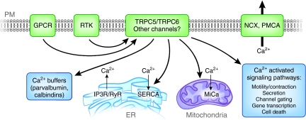
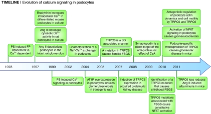
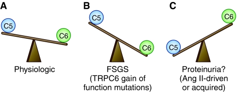
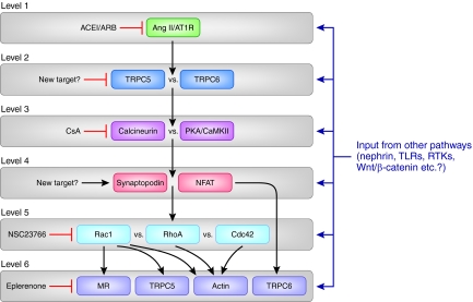
Calcium (Ca(2+)) ions are important mediators of cellular homeostasis owing to their ability to elicit a dynamic, transient, and tightly regulated range of biochemical responses. More than a decade ago, a nonselective, Ca(2+)-permeable, cationic conductance was identified in podocytes downstream of angiotensin II (Ang II) signaling, but its molecular structure remained elusive. Six years ago, transient receptor potential canonical 6 (TRPC6) mutations were found in families with hereditary FSGS, and TRPC5 and TRPC6 channels are now known as the Ca(2+) influx pathways for this previously described, nonselective, cationic current in podocytes. Ang II activation engages this Ca(2+) influx to modulate the actin cytoskeleton in podocytes. These discoveries dovetail with previously described regulation of actin dynamics by the Ca(2+)-activated phosphatase, calcineurin, and the emergence of Rho GTPases as critical regulators of podocyte function in health and disease. Understanding the interconnected signaling regulated by Ca(2+) currents offers potential new therapeutic targets and highlights the notion that synergistic therapies targeting multiple levels of biochemistry may be useful in treating proteinuric kidney disease.


Figures

Figure 1.
Figure 1.
Schematic representation of Ca2+ homeostasis in podocytes Ca2+ is a potent signaling molecule because of its ability to mediate a dynamic, dramatic, transient, and tightly regulated range of intracellular responses,, (PM: plasma membrane). Some proteins shown here have not yet been identified or studied in podocytes (calbindins, the mitochondrial uniporter or MiCa etc.), but they are likely to be present based on our understanding of calcium homeostasis in other cell types. The influx of Ca2+ is likely to be mediated by TRPC5 and TRPC6 channels, which were recorded at the single channel level in podocytes, but other influx pathways cannot be excluded. TRPC5 and TRPC6 are activated by upstream receptors such as G-protein coupled receptors (GPCR), including the AT1R, and receptor tyrosine kinases (RTK), similar to other cell types. Ca2+ is tightly regulated upon entry into the cytoplasm. Calcium homeostasis relies on the Na+-Ca2+ exchanger (NCX), which has been described in podocytes, the ATP-dependent plasma membrane Ca2+ pump (PMCA), plasma Ca2+ buffers (calbindins, parvalbumin, etc.) and internal Ca2+ stores (endoplasmic reticulum (ER), mitochondria) to maintain low cytoplasmic Ca2+ levels. When a Ca2+-permeable channel opens, whether in the plasma membrane or on a Ca2+-loaded organelle (e.g. the IP3R in the ER), Ca2+ ions flow transiently into the cytoplasm, until the homeostatic mechanisms take over once again to buffer or extrude the excess Ca2+ ions.
Figure 2.
Figure 2.
Evolution of calcium signaling in podocytes from 1978 to today.
Figure 3.
Figure 3.
Antagonistic activities of TRPC5 versus TRPC6 signaling in podocytes in health and disease: Is it a balancing act? This working model attempts to synthesize published data and underscore the areas in which future experiments are likely to enhance our understanding of TRPC signaling in podocytes. (A) Under physiologic conditions, active TRPC6 channels are more abundant on the podocyte cell membrane, as demonstrated on the single channel level, which underscores their importance for maintaining podocyte integrity, through their selective activation of RhoA., (B) TRPC6 gain of function mutations,, result in overactive TRPC6 channels, the cell is overwhelmed by TRPC6-mediated Ca2+ influx, which ultimately leads to FSGS. The observed podocyte injury may result either broadly from Ca2+ cytotoxicity and cell death or specifically from excessive RhoA-mediated contraction, for example, increased “stiffness” leading to a “broken” actin cytoskeleton, and ultimately, cell death. (C) Given the experimental evidence that (a) constitutive Rac1 activity leads to proteinuria, (b) TRPC5 activates Rac1 in podocytes, and (c) Rac1 is required for TRPC5 insertion into the plasma membrane in podocytes, it is reasonable to hypothesize that, in states of excess AngII, TRPC5/Rac1–mediated overactivity drives proteinuria. This notion generates interest in TRPC5 channels as mediators of acquired, Ang II–driven proteinuria.
Figure 4.
Figure 4.
A model for multiple signaling pathways in podocyte injury: Is a multidrug, synergistic therapy the answer to proteinuria? A synthesis of work by many groups suggests that multiple signaling levels are involved in the Ang II–mediated regulation of podocyte function in health and disease. Level 1 consists of the binding of Ang II, whose availability is limited by ACE inhibitors, to AT1Rs, which are blocked by ARBs. Subsequent activation of TRPC5 and TRPC6 channels on level 2 results in Ca2+ influx into podocytes. TRPC5-driven signaling may predominate under pathologic conditions of excess Ang II and/or Rac1 activity. TRPC5-targeted agents may therefore be a novel therapeutic approach to proteinuria. On level 3, Ca2+-activated phosphatases (calcineurin) or kinases (PKA and CamKII) battle for downstream effects on their mutual targets, synaptopodin and NFAT (level 4). Synaptopodin-preserving agents such as CsA exert their antiproteinuric effect by blocking the calcineurin-initiated degradation of synaptopodin. Stabilization of synaptopodin protein abundance or inhibition of NFAT signaling may be another therapeutic approach to proteinuria. On level 5, Rac1, RhoA, and Cdc42 battle for downstream effects on the actin cytoskeleton. Inhibition of Rac1 has an antiproteinuric effect in vivo, perhaps because of the decreased activity of mineralocorticoid receptors (MR), similar to treatment with Eplerenone (level 6). Synergistic mechanisms on level 6 include (a) the Rac1-driven insertion of TRPC5 channels in the plasma membrane (level 6), which potentiates the activity of Rac1 in a positive feedback loop, and (b) the effects of Rac1 on the actin cytoskeleton, resulting in maladaptive podocyte foot process motility, which correlates with proteinuria (level 6). RhoA and Cdc42 also affect the actin cytoskeleton (level 6). NFAT promotes TRPC6 transcription (level 6). Although AngII and AT1Rs are centrally important in podocyte Ca2+ signaling, inputs from many other pathways are likely to modulate the molecular events in each of the signaling levels, for example, signaling through the Nephrin pathway. This model also offers an explanation why the combined inhibition of several levels is necessary for effective and sustained antiproteinuric treatment (TLRs, toll-like receptors; RTKs, receptor tyrosine kinases).

References

    1. Hille B: Ionic channels of excitable membranes, Sunderland, MA, Sinauer Associates, Inc., 1992
    1. Clapham DE: Calcium signaling. Cell 131: 1047–1058, 2007 - PubMed
    1. Hodgkin AL, Huxley AF: Potassium leakage from an active nerve fibre. Nature 158: 376, 1946 - PubMed
    1. Hodgkin AL, Huxley AF: Resting and action potentials in single nerve fibres. J Physiol 104: 176–195, 1945 - PMC - PubMed
    1. Fatt P, Katz B: The effect of inhibitory nerve impulses on a crustacean muscle fibre. J Physiol 121: 374–389, 1953 - PMC - PubMed

Publication types