. 2011 Nov 11;109(11):1259-68.
doi: 10.1161/CIRCRESAHA.111.240242. Epub 2011 Oct 6.

Hydrogen sulfide as endothelium-derived hyperpolarizing factor sulfhydrates potassium channels

Affiliations

Hydrogen sulfide as endothelium-derived hyperpolarizing factor sulfhydrates potassium channels

Asif K Mustafa et al. Circ Res. .

Abstract

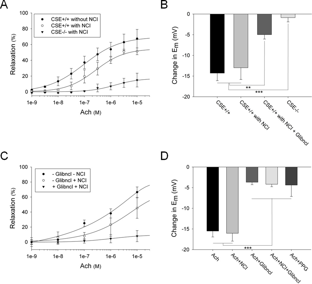
Rationale: Nitric oxide, the classic endothelium-derived relaxing factor (EDRF), acts through cyclic GMP and calcium without notably affecting membrane potential. A major component of EDRF activity derives from hyperpolarization and is termed endothelium-derived hyperpolarizing factor (EDHF). Hydrogen sulfide (H(2)S) is a prominent EDRF, since mice lacking its biosynthetic enzyme, cystathionine γ-lyase (CSE), display pronounced hypertension with deficient vasorelaxant responses to acetylcholine.

Objective: The purpose of this study was to determine if H(2)S is a major physiological EDHF.

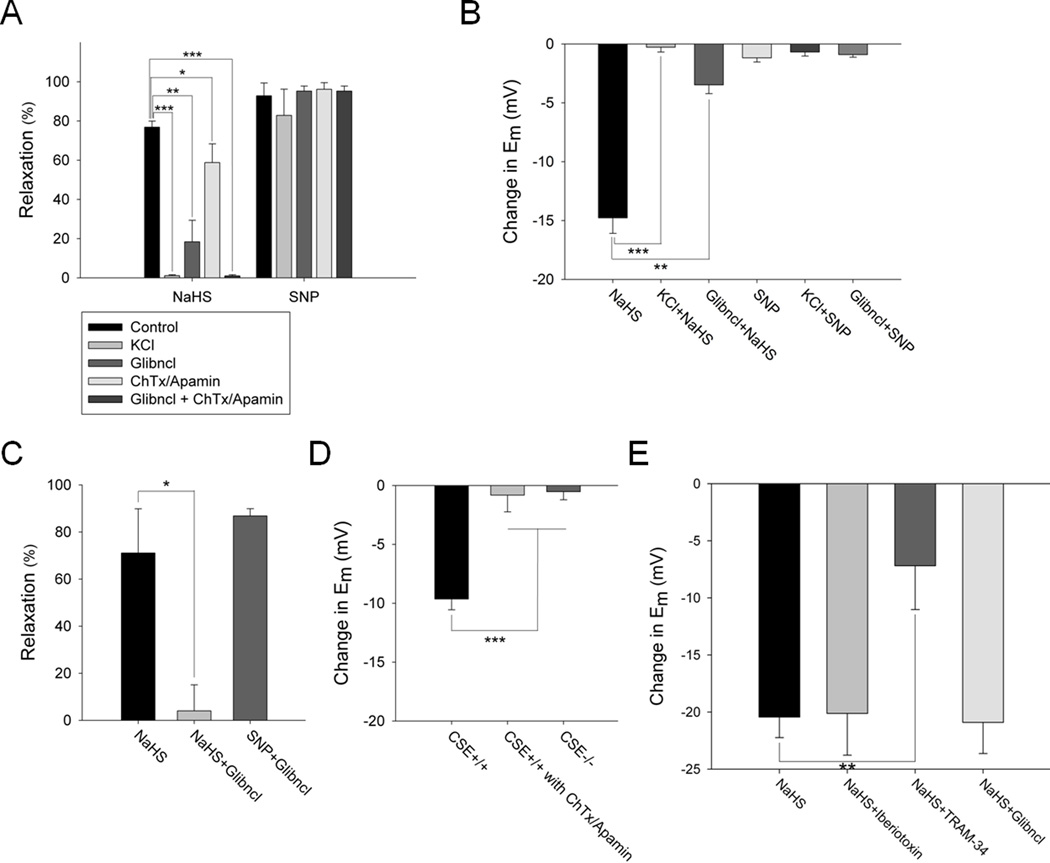
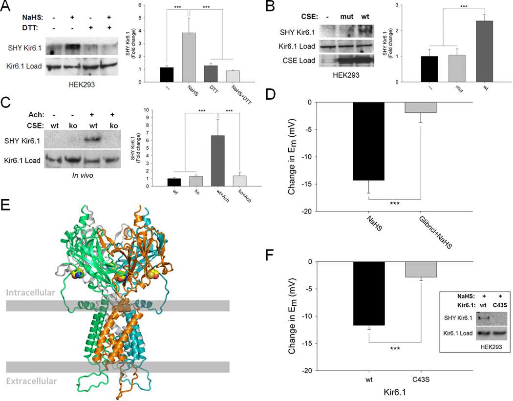
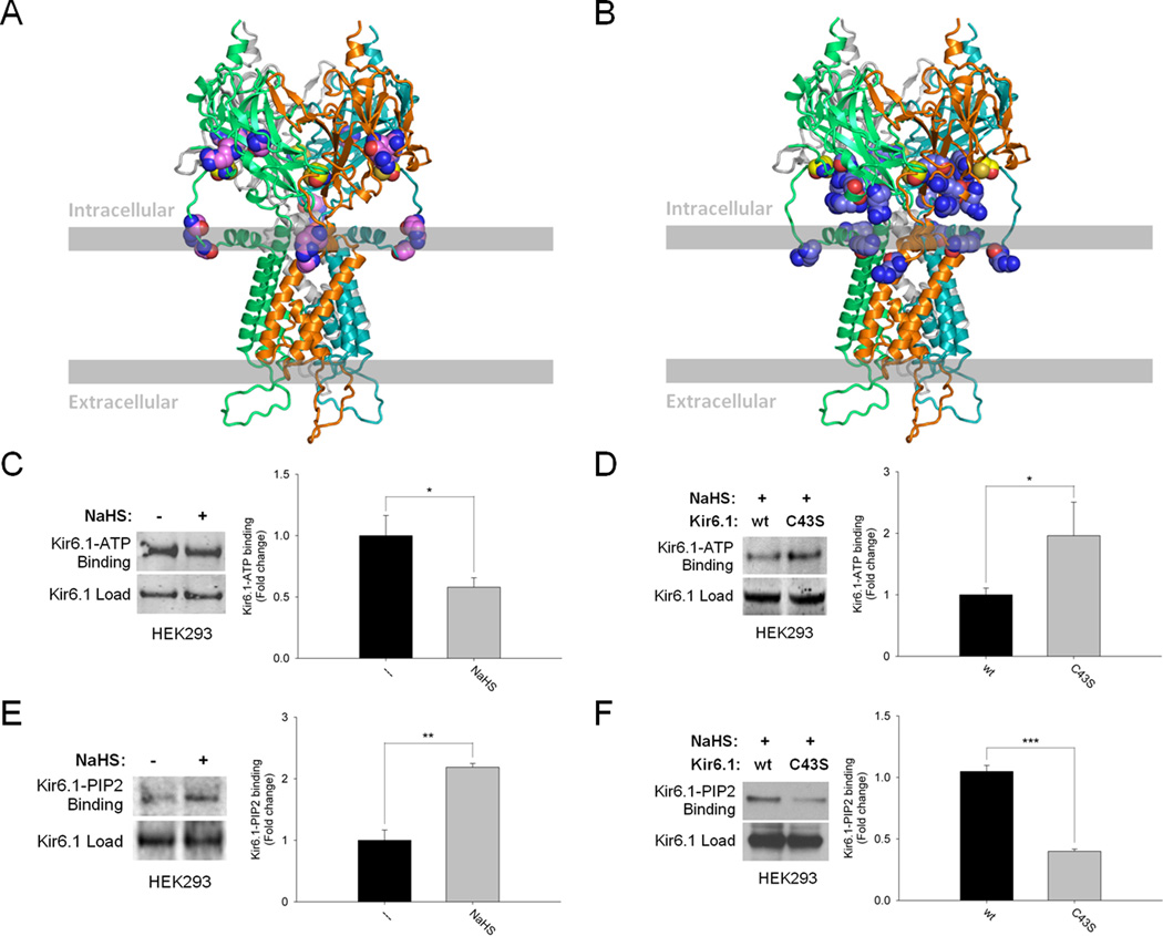
Methods and results: We now show that H(2)S is a major EDHF because in blood vessels of CSE-deleted mice, hyperpolarization is virtually abolished. H(2)S acts by covalently modifying (sulfhydrating) the ATP-sensitive potassium channel, as mutating the site of sulfhydration prevents H(2)S-elicited hyperpolarization. The endothelial intermediate conductance (IK(Ca)) and small conductance (SK(Ca)) potassium channels mediate in part the effects of H(2)S, as selective IK(Ca) and SK(Ca) channel inhibitors, charybdotoxin and apamin, inhibit glibenclamide-insensitive, H(2)S-induced vasorelaxation.

Conclusions: H(2)S is a major EDHF that causes vascular endothelial and smooth muscle cell hyperpolarization and vasorelaxation by activating the ATP-sensitive, intermediate conductance and small conductance potassium channels through cysteine S-sulfhydration. Because EDHF activity is a principal determinant of vasorelaxation in numerous vascular beds, drugs influencing H(2)S biosynthesis offer therapeutic potential.


Figures

Figure 1
Figure 1. Cholinergic vasorelaxation and hyperpolarization are significantly reduced in CSE knockout and glibenclamide treated mesenteric arteries
(A) Muscarinic cholinergic-dependent vasorelaxation of the mesenteric artery, measured using force-tension myography, is markedly diminished in CSE knockout mice compared to wild-type controls. The NOS and COX enzymes were inhibited by treatment with L-NAME (100 µM) and indomethacin (10 µM) respectively. NOS/COX inhibitors (NCI), Acetylcholine (Ach). n = 15. (B) CSE deletion almost completely abolishes the cholinergic-dependent hyperpolarization in mesenteric arteries. Treatment of wild-type mesenteric arteries with glibenclamide (5 µM) reduces the hyperpolarization by about 65%. Some of the samples were treated with L-NAME (100 µM) and indomethacin (10 µM) as indicated. The changes in membrane potential (Em) were measured with the voltage-sensitive dyes DiBAC and FLIPR. Acetylcholine was used at 10 µM. n = 24. (C) Cholinergic vasorelaxation is markedly diminished in mouse mesenteric arteries treated with glibenclamide (5 µM) in the presence of L-NAME (100 µM) and indomethacin (10 µM). n = 6. (D) Acetylcholine-mediated hyperpolarization is significantly reduced in rat mesenteric arteries treated with glibenclamide (5 µM) or propargylglycine (PPG) (10 µM). L-NAME (100 µM) and indomethacin (10 µM) do not influence membrane hyperpolarization. n = 13. All results are mean ± SEM (**p < 0.01 and ***p < 0.001).
Figure 2
Figure 2. KCl and glibenclamide markedly diminish H2S vasorelaxation and hyperpolarization in intact and endothelium-denuded mesenteric arteries
(A) H2S (100 µM) vasorelaxation of rat mesenteric arteries is completely blocked by 30 mM KCl, is reduced by 75% with glibenclamide (5 µM) alone, 25% with combination of charybdotoxin (ChTx) (1 µM) and apamin (5 µM) and almost 100% with glibenclamide and ChTx/Apamin. SNP (1 µM) vasorelaxation is not affected by any of the potassium channel inhibitors. n = 20. (B) H2S (100 µM) hyperpolarization of rat mesenteric arteries is completely blocked by 30 mM KCl and is reduced by about 75% with glibenclamide (5 µM). SNP (1 µM) does not induce hyperpolarization. n = 13. (C) H2S (100 µM) vasorelaxation in endothelium-denuded rat mesenteric artery is almost completely abolished by glibenclamide (5 µM), which fails to alter effects of SNP (1 µM). n = 6. (D) H2S hyperpolarizes endothelial cells, as seen in primary cultures of wild-type, but not CSE knockout, mouse aortic endothelial cells stimulated with acetylcholine. Treatment with ChTx/apamin completely abolishes the H2S effect. n = 10. (E) H2S hyperpolarizes human aortic endothelial cells (HAEC) treated with Iberiotoxin (0.5 µM) or glibenclamide (0.1 µM), but not TRAM-34 (10 nM). n = 8. All results are mean ± SEM (*p < 0.05, **p < 0.01 and ***p < 0.001).
Figure 3
Figure 3. Physiologic sulfhydration of Kir 6.1-cysteine-43 activates the channel causing hyperpolarization
(A) H2S (100 µM) sulfhydrates (SHY) Kir 6.1 overexpressed in HEK293 cells, an effect reversed by DTT (1 mM). n = 4. (B) Kir 6.1 is basally sulfhydrated in cells overexpressing catalytically-active wild-type (wt) CSE but not in cells lacking CSE or containing catalytically-inactive mutant CSE (mut). n = 4. (C) Cholinergic stimulation of mouse aorta enhances sulfhydration of Kir 6.1 in wild-type but not CSE knockout (ko) mice. n = 3. (D) H2S (100 µM)-elicited hyperpolarization in HEK293 cells overexpressing Kir 6.1 is substantially reduced by glibenclamide (5 µM). n = 7. (E) Model of Kir 6.1 homotetramer based on the established structure of Kir 3.1 with surface residue cysteine-43 highlighted in yellow. (F) H2S (300 µM)-mediated sulfhydration (inset) and hyperpolarization are absent in HEK293 cells overexpressing C43S mutant Kir 6.1. n = 12. Quantitative densitometric analysis is also shown for Figure 3A–C. All results are mean ± SEM (***p < 0.001).
Figure 4
Figure 4. Sulfhydration augments ATP-sensitive potassium channel activity by reducing Kir 6.1-ATP binding and enhancing Kir 6.1-PIP2 binding
(A) Model of Kir 6.1 with cysteine-43 highlighted in yellow as well as ATP interacting residues (R51, G54, R195 and R211) highlighted in violet., (B) Model of Kir 6.1 with cysteine-43 highlighted in yellow and PIP2 interacting residues (R55, K68, R186, R187, R216 and R310) in stale blue., (C) Sulfhydration of Kir 6.1 in HEK293 cells reduces its interaction with ATP. n = 4. (D) Kir 6.1-ATP interaction is substantially enhanced in H2S (100 µM)-treated HEK293 cells overexpressing Kir 6.1-C43S mutant. n = 3. (E) Sulfhydration of Kir 6.1 in HEK293 cells markedly augments its binding to PIP2. n = 3. (F) Kir 6.1-PIP2 interaction is significantly reduced in H2S (100 µM)-treated HEK293 cells overexpressing Kir 6.1-C43S mutant. n = 4. Quantitative densitometric analysis is also shown for Figure 4C–F. All results are mean ± SEM (*p < 0.05, **p < 0.01 and ***p < 0.001).

Comment in

References

    1. Murad F. Shattuck Lecture. Nitric oxide and cyclic GMP in cell signaling and drug development. N Engl J Med. 2006 Nov 9;355(19):2003–2011. - PubMed
    1. Furchgott RF, Zawadzki JV. The obligatory role of endothelial cells in the relaxation of arterial smooth muscle by acetylcholine. Nature. 1980 Nov 27;288(5789):373–376. - PubMed
    1. Ignarro LJ, Buga GM, Wood KS, Byrns RE, Chaudhuri G. Endothelium-derived relaxing factor produced and released from artery and vein is nitric oxide. Proc Natl Acad Sci U S A. 1987 Dec;84(24):9265–9269. - PMC - PubMed
    1. Moncada S, Higgs EA. The discovery of nitric oxide and its role in vascular biology. Br J Pharmacol. 2006 Jan;147 Suppl 1:S193–S201. - PMC - PubMed
    1. Feletou M, Vanhoutte PM. Endothelium-dependent hyperpolarizations: past beliefs and present facts. Ann Med. 2007;39(7):495–516. - PubMed

Publication types

MeSH terms