. 2011:6:1793-800.
doi: 10.2147/IJN.S23638. Epub 2011 Aug 26.

Metabolic pathway and distribution of superparamagnetic iron oxide nanoparticles: in vivo study

Affiliations

Metabolic pathway and distribution of superparamagnetic iron oxide nanoparticles: in vivo study

Eva K Schlachter et al. Int J Nanomedicine. 2011.

Abstract

Background: Experimental tissue fusion benefits from the selective heating of superparamagnetic iron oxide nanoparticles (SPIONs) under high frequency irradiation. However, the metabolic pathways of SPIONs for tissue fusion remain unknown. Hence, the goal of this in vivo study was to analyze the distribution of SPIONs in different organs by means of magnetic resonance imaging (MRI) and histological analysis after a SPION-containing patch implantation.

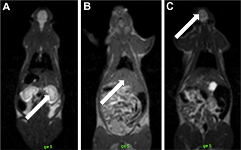
Methods: SPION-containing patches were implanted in rats. Three animal groups were studied histologically over six months. Degradation assessment of the SPION-albumin patch was performed in vivo using MRI for iron content localization and biodistribution.

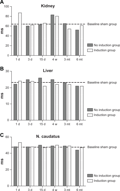
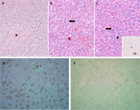
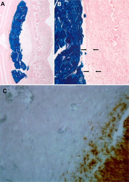
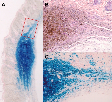
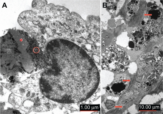
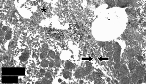
Results: No SPION degradation or accumulation into the reticuloendothelial system was detected by MRI, MRI relaxometry, or histology, outside the area of the implantation patch. Concentrations from 0.01 μg/mL to 25 μg/mL were found to be hyperintense in T1-like gradient echo sequences. The best differentiation of concentrations was found in T2 relaxometry, susceptibility-sensitive gradient echo sequences, and in high repetition time T2 images. Qualitative and semiquantitative visualization of small concentrations and accumulation of SPIONs by MRI are feasible. In histological liver samples, Kupffer cells were significantly correlated with postimplantation time, but no differences were observed between sham-treated and induction/no induction groups. Transmission electron microscopy showed local uptake of SPIONs in macrophages and cells of the reticuloendothelial system. Apoptosis staining using caspase showed no increased toxicity compared with sham-treated tissue. Implanted SPION patches were relatively inert with slow, progressive local degradation over the six-month period. No distant structural alterations in the studied tissue could be observed.

Conclusion: Systemic bioavailability may play a role in specific SPION implant toxicity and therefore the local degradation process is a further aspect to be assessed in future studies.

Keywords: distribution; metabolism; superparamagnetic iron oxide nanoparticles.


Figures

Figure 1
Figure 1
Three full-body magnetic resonance scans in a rat. After choosing a suitable slice position for each rat, standardized regions of interest are placed in homogenous parts of (A) kidney, (B) liver, and (C) nucleus caudatus (marked with arrows) to measure and compare T2 relaxation times.
Figure 2
Figure 2
Comparison of the half-life values in milliseconds of the decay curves in all tested (A) livers, (B) kidneys, and (C) nucleus caudatus of animals with implanted patches. For better visualization, the mean values for the sham-operated group is marked as a bar. The values show no systematic variation, regardless of whether a sham-operated, induction, or no induction group. Variation of values in the kidney is probably a normal phenomenon caused by nonhomogeneous regions of interest regarding the constantly changing water content. The same amplitude of variation can be found in the sham group.
Figure 3
Figure 3
Conventional light microscopic histology of representative rat liver tissue removed (A) three days and (B) six months after implantation of superparamagnetic iron oxide nanoparticles. (C) Liver tissue of a sham-treated animal after six months. All visual fields include a central vein. Neither iron storage in hepatocytes nor fibrosis is seen. Arrows in (B) and (C) point to minute iron deposits in a Kupffer cell flanking a sinusoid. This chance finding is due to age-related increase of Kupffer cells and is therefore less likely to be encountered in young animals. (A) Iron granules are readily distinguished from lipofuscin pigment using Prussian Blue staining (inset in C). Slides not labeled otherwise represent hematoxylin and eosin staining; original magnification 200×. Representative section of rat liver using active caspase-3 antibody staining at three days (D) 40× and cleaved caspase-3 antibody staining at six months (E) 40× demonstrated no apoptotic activity. Sham caspase is not shown because no difference was detected.
Figure 4
Figure 4
Histological findings of the superparamagnetic iron oxide nanoparticle-albumin complex three days after implantation and heat treatment in an electromagnetic field for 60 seconds. (A) Scanning magnification shows implant smoothly accommodated along the cleavage plane of subcutaneous fascia and axial musculature with no significant space-occupying effect. (B) Detailed view of implant/ tissue interface indicates this to consist of a narrow rim of fibrinoid exudate (arrows) surrounded by granulation tissue. Prussian Blue staining; original magnification (A) 15× and B 100×. In (C), active caspase-3 antibody staining shows no apoptotic activity around the implant (40×). Sham not shown because no difference was detected.
Figure 5
Figure 5
Histological aspect of superparamagnetic iron oxide nanoparticle-albumin complex in situ six months after implantation and heating procedure. (A) At scanning magnification of Prussian Blue-stained section, centrifugal fading rather than sharp circumscription of implant contours is seen (especially as opposed to Figure 7A). (B) and (C) represent consecutive section planes of boxed area in (A) to indicate gradual dissolution of implant iron content by macrophages. In parallel, there is fibroblastic ingrowth. Note absence of significant foreign body reaction. (A) and (C) Prussian Blue staining, (B) hematoxylin and eosin staining; original magnifications (A) 15× and (B) and (C) 200×.
Figure 6
Figure 6
Ultrastructural aspect of implant/tissue interaction as seen on transmission electron microscopy. (A) Detailed view of early phase of implant degradation showing ongoing engulfment of the superparamagnetic iron oxide nanoparticles-albumin complex by activated monocytes. Heterogeneous electron density of implant material is seen, with electron-lucent zones corresponding to coagulated albumin (*) whereas dense granules represent iron nanoparticles (○). (B) Organizing phase of implant scavenging is characterized by phagocytic cells replete with electron-dense iron particles. Note intercellular deposition of connective tissue fibers (arrows). Original magnification is indicated by scale bars.
Figure 7
Figure 7
Ultrastructural aspect of liver from an implant-bearing animal in the late phase of observation period. *Transected biliary canalicule flanked by two hepatocytes, the cell borders of which (arrows) are not readily identified due to poor specimen preservation. Regular aspect of mitochondria is a good indicator of lack of metabolic stress, as frequently seen in storage disorders. Original magnification is indicated by scale bar.

References

    1. Babincova M, Leszczynska D, Sourivong P, Babinec P. Selective treatment of neoplastic cells using ferritin-mediated electromagnetic hyperthermia. Med Hypotheses. 2000;54(2):177–179. - PubMed
    1. Berry CC. Progress in functionalization of magnetic nanoparticles for applications in biomedicine. J Phys D Appl Phys. 2009;42(22):224003.
    1. Bregy A, Kohler A, Steitz B, et al. Electromagnetic tissue fusion using superparamagnetic iron oxide nanoparticles: First experience with rabbit aorta. The Open Surgery Journal. 2008;(2):3–9.
    1. van Landeghem FK, Maier-Hauff K, Jordan A, et al. Post-mortem studies in glioblastoma patients treated with thermotherapy using magnetic nanoparticles. Biomaterials. 2009;30(1):52–57. - PubMed
    1. Lacava LM, Lacava ZG, Da Silva MF, et al. Magnetic resonance of a dextran-coated magnetic fluid intravenously administered in mice. Biophys J. 2001;80(5):2483–2486. - PMC - PubMed

Publication types

LinkOut - more resources