. 2011 Sep;7(9):e1002259.
doi: 10.1371/journal.ppat.1002259. Epub 2011 Sep 29.

Monalysin, a novel ß-pore-forming toxin from the Drosophila pathogen Pseudomonas entomophila, contributes to host intestinal damage and lethality

Affiliations

Monalysin, a novel ß-pore-forming toxin from the Drosophila pathogen Pseudomonas entomophila, contributes to host intestinal damage and lethality

Onya Opota et al. PLoS Pathog. 2011 Sep.

Abstract

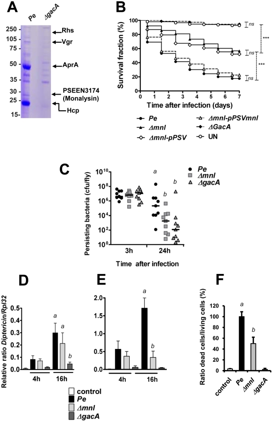
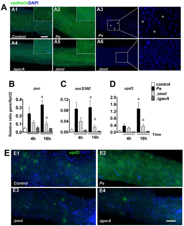
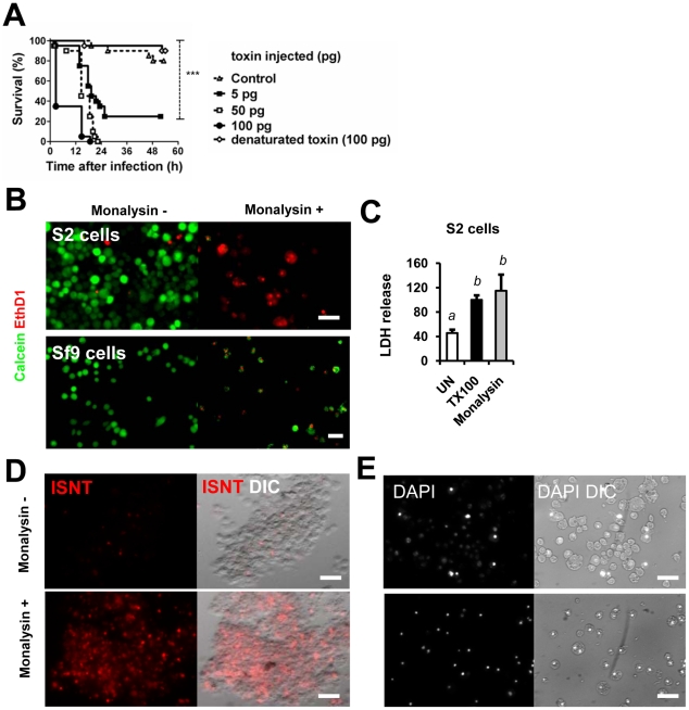
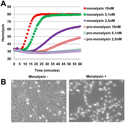
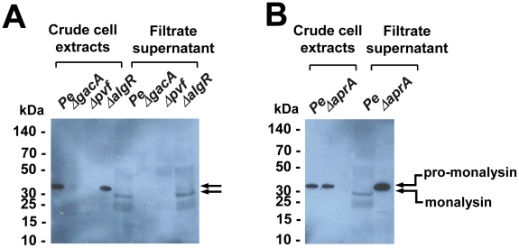
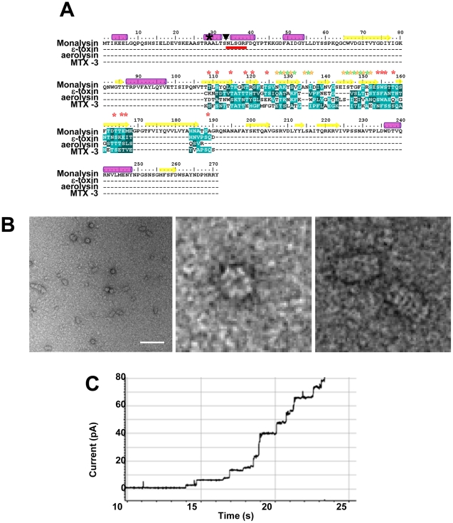
Pseudomonas entomophila is an entomopathogenic bacterium that infects and kills Drosophila. P. entomophila pathogenicity is linked to its ability to cause irreversible damages to the Drosophila gut, preventing epithelium renewal and repair. Here we report the identification of a novel pore-forming toxin (PFT), Monalysin, which contributes to the virulence of P. entomophila against Drosophila. Our data show that Monalysin requires N-terminal cleavage to become fully active, forms oligomers in vitro, and induces pore-formation in artificial lipid membranes. The prediction of the secondary structure of the membrane-spanning domain indicates that Monalysin is a PFT of the ß-type. The expression of Monalysin is regulated by both the GacS/GacA two-component system and the Pvf regulator, two signaling systems that control P. entomophila pathogenicity. In addition, AprA, a metallo-protease secreted by P. entomophila, can induce the rapid cleavage of pro-Monalysin into its active form. Reduced cell death is observed upon infection with a mutant deficient in Monalysin production showing that Monalysin plays a role in P. entomophila ability to induce intestinal cell damages, which is consistent with its activity as a PFT. Our study together with the well-established action of Bacillus thuringiensis Cry toxins suggests that production of PFTs is a common strategy of entomopathogens to disrupt insect gut homeostasis.


Conflict of interest statement

The authors have declared that no competing interests exist.

Figures

Figure 1
Figure 1. PSEEN3174 encodes a secreted protein, Monalysin, required for P. entomophila virulence.
(A) SDS-PAGE analysis of culture supernatants from wild type P. entomophila and the ΔgacA derivative. Proteins extracted from culture supernatants were loaded on a SDS-PAGE and stained with coomassie blue. The nature of proteins identified by MALDI-TOF analysis of tryptic fragments is shown on the right. (B) Survival analysis of wild-type Oregon adult flies following infection by feeding with the P. entomophila wild-type strain (Pe), the gacA-deficient strain (ΔgacA), the mnl deficient strain (Δmnl) the mnl-deficient strain carrying a plasmid expressing a wild-type copy of the monalysin gene (Δmnl-pPSVmnl), or carrying the plasmid pPSV35 without any insert (Δmnl-pPSV). UN: unchallenged. The Kaplan-Meier log rank test was used to determine statistical significance. Dashed brackets represent the significance between the different infections (***: p<0.001, ns: not significant). (C) Bacterial persistence in wild-type Oregon flies as the number of colony-forming-unit (cfu) per fly. After infection by the P. entomophila wild-type strain (Pe), the gacA-deficient strain (ΔgacA), the mnl deficient strain (Δmnl), the number of cfu per fly was determined at the indicated time point. (D and E )Time-course analysis of Diptericin expression measured by RT-qPCR in (D) guts (local) or (E) whole flies (corresponding to the systemic fat body expression). (F) Cell death quantification using acridine orange staining. Results represent the percentage of dead cells (acridine orange positive nuclei) in the midguts of flies infected for 16 hours with the indicated bacterial strains. Results represent the average of four independent experiments. Statistical analysis was performed using a Wilcoxon test, and different letters indicate significantly different values (P<0.05).
Figure 2
Figure 2. Monalysin contributes to P. entomophila-inflicted damage to the Drosophila gut.
(A) Expression of the marker of adherens junction Cadherin-GFP 16 hours after infection with lethal doses of the indicated bacteria. Ingestion of wild type P. entomophila disrupts the pattern of Cadherin-GFP. A3 and A6 show DAPI of A2 and A5 respectively. The symbol (*) marks regions where DAPI staining is absent. (B–D) Analysis of puckered (puc), unpaired3 (upd3), and socs36E expression measured by RT-qPCR in guts of infected flies. Statistical analysis was performed using a Wilcoxon test and letters indicate significantly different values (P<0.05). (E) Expression of the upd3-Gal4, UAS-GFP reporter in (E1) unchallenged flies or( E2–E4) 4 hours after infections with a sublethal dose of bacteria (OD600 = 10). In contrast to the wild type P. entomophila strain (E2), the Δmnl (E3) and the ΔgacA (E4) strains were unable to elicit upd3-GFP expression. Scale bars represent 50 µm.
Figure 3
Figure 3. Monalysin encodes a cytotoxic protein secreted by P. entomophila.
(A) Survival analysis of wild-type Oregon adult flies after injection of various quantities of Monalysin or heat-inactivated (denaturated) Monalysin. (B and C) Cytotoxic effect of Monalysin on insect culture cell line S2. (B) Drosophila S2 cells and Spodoptera frugiperda Sf9 cells were treated with Monalysin (Final concentration = 100nM) and stained with a live-dead viability reagent. Living cells are stained in green with Calcein while dead cells are stained in red with Ethidium homodimer 1 (EthD1, red). (C) The loss of viability was quantified by measuring the release of lactate dehydrogenase (LDH) from S2 cells. (D) DNA fragmentation in S2 cells was monitored by ISNT (in situ nick translation). (E) Chromatin condensation on untreated and Monalysin treated S2 cells was examined by DAPI staining. Phase-contrast and fluorescence views of the same microscopic fields are shown. (−) untreated cells, (+)  =  cells treated with Monalysin 100nM.
Figure 4
Figure 4. Monalysin hemolytic activity and cytotoxicity towards mammalian cells.
(A) Hemolytic activity was measured with pro and matured Monalysin incubated with red blood cells. The mature Monalysin was obtained by limited trypsinolysis of a fresh extract of recombinant Pro-Monalysin. (B) Phase contrast microscopy of Hela cells untreated or treated with Monalysin 100 nM for 24h. Cells were shrinking and displayed irreversible loss of adherence.
Figure 5
Figure 5. Regulation and processing of Monalysin.
(A) Western-blot analysis of proteins from crude cell extracts or filtrate supernatants shows that Monalysin was not produced in gacA and pvf mutants, but was produced in the algR mutant. (B) Western-blot analysis of bacterial crude cell extracts and filtrated supernatants of Pe wt and ΔaprA shows that pro-monalysin is not processed in the supernatant of an AprA mutant. The stronger signal in the AprA mutant lane is due to the fact that the serum recognized better the pro-monalysin than the monalysin (see Figure S5).
Figure 6
Figure 6. Monalysin is a ß pore-forming toxin.
(A) Protein sequence analysis of Monalysin reveals an internal domain with amphipathic patches flanked by serine- and threonine-rich sequences that shares similarities with the membrane-spanning domain of ß-PFT (ε-toxin from Clostridium perfringiens, aerolysin from Aeromonas hydrophila and MTX-3 from Bacillus sphaericus). The multiple sequence alignment reveals the presence in P. entomophila Monalysin of putative membrane exposed residues (Yellow stars), solvent-exposed residues (green stars), and serine and threonine residues (red stars). A black star (★) shows the first amino acid detected by MALDI-TOF analysis of tryptic fragment of the recombinant Monalysin. The N-terminal residues of the mature Monalysin, identified by Edman sequencing, present in P. entomophila supernatant are underlined in red; a triangle (▾) indicates the potential cleavage site of pro-monalysin deduced from the N-terminal Edman sequencing. Purple cylinders indicate predicted α-helixes and yellow arrows indicate predicted β-sheets. (B) Scanning Electron micrographs show that Monalysin forms circular-like structures (top view) and barrel-like aggregates (side view). Scale bar represents 100 nm. (C) Monalysin (5 µg.ml) is able to form pores in a planar lipid bilayer.

References

    1. Sansonetti PJ. War and peace at mucosal surfaces. Nat Rev Immunol. 2004;4:953–964. - PubMed
    1. Schneider DS, Ayres JS. Two ways to survive infection: what resistance and tolerance can teach us about treating infectious diseases. Nat Rev Immunol. 2008;8:889–895. - PMC - PubMed
    1. Hooper LV, Macpherson AJ. Immune adaptations that maintain homeostasis with the intestinal microbiota. Nat Rev Immunol. 2010;10:159–169. - PubMed
    1. Vodovar N, Vinals M, Liehl P, Basset A, Degrouard J, et al. Drosophila host defense after oral infection by an entomopathogenic Pseudomonas species. Proc Natl Acad Sci U S A. 2005;102:11414–11419. - PMC - PubMed
    1. Vodovar N, Vallenet D, Cruveiller S, Rouy Z, Barbe V, et al. Complete genome sequence of the entomopathogenic and metabolically versatile soil bacterium Pseudomonas entomophila. Nat Biotechnol. 2006;24:673–679. - PubMed

Publication types

MeSH terms

LinkOut - more resources