. 2011;6(9):e24992.
doi: 10.1371/journal.pone.0024992. Epub 2011 Sep 27.

Intravascular food reward

Affiliations

Intravascular food reward

Albino J Oliveira-Maia et al. PLoS One. 2011.

Abstract

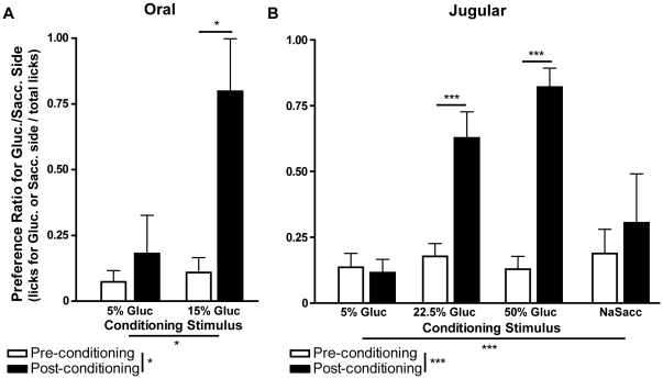
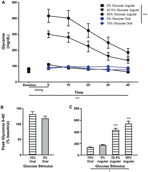
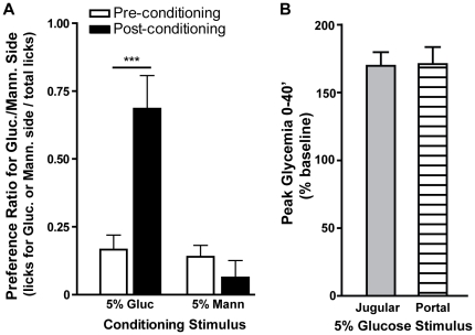
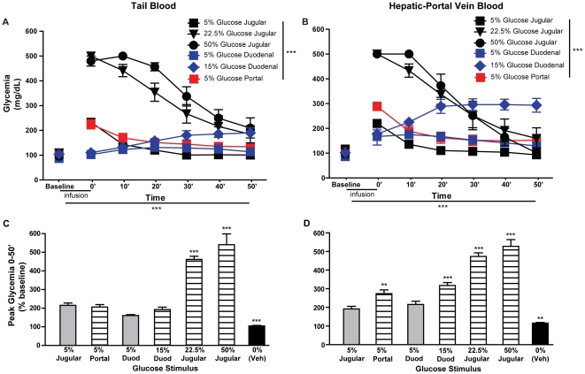
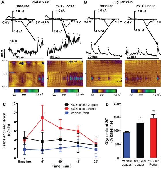
Consumption of calorie-containing sugars elicits appetitive behavioral responses and dopamine release in the ventral striatum, even in the absence of sweet-taste transduction machinery. However, it is unclear if such reward-related postingestive effects reflect preabsorptive or postabsorptive events. In support of the importance of postabsorptive glucose detection, we found that, in rat behavioral tests, high concentration glucose solutions administered in the jugular vein were sufficient to condition a side-bias. Additionally, a lower concentration glucose solution conditioned robust behavioral responses when administered in the hepatic-portal, but not the jugular vein. Furthermore, enteric administration of glucose at a concentration that is sufficient to elicit behavioral conditioning resulted in a glycemic profile similar to that observed after administration of the low concentration glucose solution in the hepatic-portal, but not jugular vein. Finally using fast-scan cyclic voltammetry we found that, in accordance with behavioral findings, a low concentration glucose solution caused an increase in spontaneous dopamine release events in the nucleus accumbens shell when administered in the hepatic-portal, but not the jugular vein. These findings demonstrate that the postabsorptive effects of glucose are sufficient for the postingestive behavioral and dopaminergic reward-related responses that result from sugar consumption. Furthermore, glycemia levels in the hepatic-portal venous system contribute more significantly for this effect than systemic glycemia, arguing for the participation of an intra-abdominal visceral sensor for glucose.


Conflict of interest statement

Competing Interests: The authors have declared that no competing interests exist.

Figures

Figure 1
Figure 1. Jugular vein administration of glucose is sufficient to condition side-bias reversal.
A. In animals conditioned with 15%, but not 5% glucose, delivered orally, there was a statistically significant reversal of original side-bias. B. In animals conditioned with stimuli administered in the JV, only those exposed to 22.5% and 50% glucose, but not 5% glucose or 3.16% NaSacc (to control for intravascular taste), reversed side-bias after conditioning. *p<0.05, **p<0.001, ***p<0.0001.
Figure 2
Figure 2. Jugular vein infusion of glucose stimuli that condition side-bias reversal also lead to extreme tail-blood hyperglycemia.
A. In awake animals, glycemia (mg/dL) was measured from tail blood at the start (baseline), end (0′) and at 10 minute intervals after (10′–40′) behavioral sessions where one of the glucose solutions used for conditioning was administered. Significant overall effects were found for stimulus (5% vs. 22.5% vs. 50% JV glucose vs. 5% vs. 15% oral glucose; F = 25.5, p<0.0001), time (baseline vs. 0′ vs. 10′ vs. 20′ vs. 30′ vs. 40′; F = 38, p<0.0001) and the interaction between these factors (F = 12.6, p<0.0001; repeated-measures two-way ANOVA). Given this interaction, data was analyzed separately for each stimulus, showing a significant effect for time in all cases (JV 5% glucose, F = 14.5, p<0.0001; JV 22.5% glucose, F = 20, p<0.0001; JV 50% glucose, F = 28, p<0.0001; oral 15% glucose, F = 3.2, p = 0.036) with the exception of oral 5% glucose (F = 2.7, p = 0.087; repeated-measures one-way ANOVA). Furthermore, when glycemia at each time-point was compared to the respective baseline measure, significant effects were found at several time-points for JV 5% glucose (0′ and 10′), JV 22.5% glucose (0′, 10′, 20′ and 30′), JV 50% glucose (0′, 10′, 20′, 30′, and 40′), oral 15% glucose (10′, 20′ and 30′), but not for 5% oral glucose (post-hoc Bonferroni t-tests, see Table S1 for details). B–C. Peak relative glycemia (% of baseline) was compared after administration of glucose stimuli previously shown to be effective to condition side-bias reversal (hatched bars) and others that failed to do so (gray bars). We did not find significant differences between animals that consumed 5% or 15% glucose orally (B). Furthermore, glycemia after oral delivery of 15% glucose was significantly lower relative to that found after other stimuli that induced side-bias reversal (22.5% and 50%), but did not differ relative to 5% glucose, that did not condition such reversal (C). *p<0.05, ***p<0.0001.
Figure 3
Figure 3. Hepatic-portal vein administration of glucose conditions side-bias reversal at lower concentrations than those needed with jugular vein administration.
A. In animals conditioned with 5% glucose, but not 5% mannitol, administered in the HPV, we found a significant reversal of original side-bias. B. In awake animals, peak tail blood glycemia (% of baseline) after HPV 5% glucose, that conditioned side-bias reversal (hatched bar), was not significantly different from that observed after JV administration of 5% glucose, that failed to condition these behavioral effects (gray bar). ***p<0.0001.
Figure 4
Figure 4. Increases in hepatic-portal vein blood glycemia parallel the capability of glucose stimuli to condition side-bias reversal.
Tail and HPV blood glycemia was measured in anesthetized rats at the start (baseline) and several time-points after (0′–50′) perfusion of vehicle (duodenal water, JV saline or HPV saline) or one of the different glucose solutions used for conditioning (duodenal 5% or 15% glucose, JV 5%, 22.5% or 50% glucose or HPV 5% glucose). A. For tail blood glycemia, measurements after glucose injections, a significant overall effect was found for time, stimulus and the interaction between these factors (p<0.0001 for all; repeated-measures two-way ANOVA). Further comparisons were performed relative to JV 5% glucose, considered as a control stimulus that did not condition side-bias reversal, revealing several time-points after JV 22.5% and 50% and duodenal 15% glucose where glycemia was significantly elevated (see Table S2 for details). B. In terms of HPV blood glycemia, measurements after glucose injections, a significant overall effect was found for time, stimulus and their interaction (p<0.0001 for all; repeated-measures two-way ANOVA). Again, significant elevations of glycemia where found at several time-points after JV 22.5% and 50% and duodenal 15% glucose, when compared to those verified after JV 5% glucose (see Table S2 for details). C–D. Peak relative glycemia (% of baseline) was also compared after administration of vehicle (black bars), glucose stimuli previously shown to be effective to condition side-bias reversal (hatched bars) and others that failed to do so (gray bars). Peak glycemia for each stimulus was again compared to that after to JV 5% glucose, considered as a control stimulus that did not condition side-bias reversal and differences were parallel to the behavioral effects of each stimulus for HPV (D) but not tail (C) blood measurements. **p<0.001, ***p<0.0001.
Figure 5
Figure 5. Nucleus accumbens dopamine transient frequency increases after injection of glucose in the hepatic-portal vein.
Fast-scan cyclic voltammetry was used to identify spontaneous dopamine release events (transients) in the nucleus accumbens shell of anesthetized rats. Measurements were conducted for a baseline period, and also during and after infusion of 5% glucose in the JV (n = 4) or HPV (n = 4), or vehicle in the latter (n = 4). A. This panel shows example measurements in one animal before (figures on the left) and after (figures on the right) onset of 5% glucose infusion in the HPV. The two dimensional color plots at the bottom of the panel show the voltammetric current (encoded in color, nA) plotted against the applied potential on the Y axis (V) and the acquisition time on the X axis (sec.). The green spots at 0.62 V (the peak oxidation potential for dopamine) in the lower half of the plots represent dopamine oxidation at the surface of the carbon-fiber microelectrode. The current at the peak dopamine oxidation potentials was converted to dopamine concentration following post-calibration (see Methods) and is displayed above the color plots, approximately aligned with them in time. The panels near the top of the figure are cyclic voltammograms of the peaks indicated by arrows that were obtained by subtracting baseline voltammograms just prior to onset of the transient. Dopamine transients were considered as such when they had voltammograms that were significantly correlated (r>0.86) with those of a dopamine template elicited previously by electrical stimulation of the ventral tegmental area. The peaks that were thus classified as dopamine are indicated by asterisks. B. This panel shows an example measurement as was described above (A) but for JV, rather than HPV administration of 5% glucose. Note that the evoked activity is much less. C. Here we show dopamine transient frequency prior to infusion in each of the three groups (baseline) and in 5 min bins following infusion onset. Data were analyzed using two-way repeated measures ANOVA (see text) and post-hoc analysis indicated that, when compared to the respective time period for HPV vehicle administration, transient frequency in the HPV glucose group was significantly higher in the first 5 minutes after infusion onset (t = 3.1, p<0.05) but not in the remaining time periods (t<1.9, p>0.05 for all), or at any point in the JV glucose group (t<1.1, p>0.05 for all; post-hoc Bonferroni t-tests). D. In the same rats, tail blood glycemia was also measured prior to and 20 minutes after infusion of 5% glucose in the JV (n = 3) or HPV (n = 3), or vehicle in the latter (n = 4). No differences for absolute glycemia (mg/dL) were found between groups at baseline (PV vehicle - 157±7, JV 5% glucose - 169±22, HPV 5% glucose - 135±14; F = 1.4, p = 0.31, repeated-measures one-way ANOVA; t<1.7, p>0.05 for all pair-wise comparisons, post-hoc Bonferroni t-tests; data not shown in figure). However, as expected, an overall significant difference was found for relative glycemia (% baseline) at 20′ (F = 14.6, p = 0.0032, repeated-measures one-way ANOVA), with significant differences between glycemia after HPV vehicle (93±4) and that after JV 5% glucose (129±6; t = 3.5, p<0.05) and HPV 5% glucose (147±12; t = 5.2, p<0.01). Importantly, no differences were found between values after each of the two glucose stimuli (t = 1.6, p>0.05, post-hoc Bonferroni t-tests). *p<0.05, **p<0.001.

References

    1. Sclafani A. Oral and postoral determinants of food reward. Physiol Behav. 2004;81:773–779. - PubMed
    1. de Araujo IE, Oliveira-Maia AJ, Sotnikova TD, Gainetdinov RR, Caron MG, et al. Food reward in the absence of taste receptor signaling. Neuron. 2008;57:930–941. - PubMed
    1. Frank GK, Oberndorfer TA, Simmons AN, Paulus MP, Fudge JL, et al. Sucrose activates human taste pathways differently from artificial sweetener. Neuroimage. 2008;39:1559–1569. - PubMed
    1. Touzani K, Bodnar R, Sclafani A. Activation of dopamine D1-like receptors in nucleus accumbens is critical for the acquisition, but not the expression, of nutrient-conditioned flavor preferences in rats. Eur J Neurosci. 2008;27:1525–1533. - PubMed
    1. Ackroff K, Yiin YM, Sclafani A. Post-oral infusion sites that support glucose-conditioned flavor preferences in rats. Physiol Behav. 2010;99:402–411. - PMC - PubMed

Publication types