. 2011;6(9):e25450.
doi: 10.1371/journal.pone.0025450. Epub 2011 Sep 30.

Activation of WNT/β-catenin signaling in pulmonary fibroblasts by TGF-β₁ is increased in chronic obstructive pulmonary disease

Affiliations

Activation of WNT/β-catenin signaling in pulmonary fibroblasts by TGF-β₁ is increased in chronic obstructive pulmonary disease

Hoeke A Baarsma et al. PLoS One. 2011.

Abstract

Background: Chronic obstructive pulmonary disease (COPD) is characterized by abnormal extracellular matrix (ECM) turnover. Recently, activation of the WNT/β-catenin pathway has been associated with abnormal ECM turnover in various chronic diseases. We determined WNT-pathway gene expression in pulmonary fibroblasts of individuals with and without COPD and disentangled the role of β-catenin in fibroblast phenotype and function.

Methods: We assessed the expression of WNT-pathway genes and the functional role of β-catenin, using MRC-5 human lung fibroblasts and primary pulmonary fibroblasts of individuals with and without COPD.

Results: Pulmonary fibroblasts expressed mRNA of genes required for WNT signaling. Stimulation of fibroblasts with TGF-β₁, a growth factor important in COPD pathogenesis, induced WNT-5B, FZD₈, DVL3 and β-catenin mRNA expression. The induction of WNT-5B, FZD₆, FZD₈ and DVL3 mRNA by TGF-β₁ was higher in fibroblasts of individuals with COPD than without COPD, whilst basal expression was similar. Accordingly, TGF-β₁ activated β-catenin signaling, as shown by an increase in transcriptionally active and total β-catenin protein expression. Furthermore, TGF-β₁induced the expression of collagen1α1, α-sm-actin and fibronectin, which was attenuated by β-catenin specific siRNA and by pharmacological inhibition of β-catenin, whereas the TGF-β₁-induced expression of PAI-1 was not affected. The induction of transcriptionally active β-catenin and subsequent fibronectin deposition induced by TGF-β₁ were enhanced in pulmonary fibroblasts from individuals with COPD.

Conclusions: β-catenin signaling contributes to ECM production by pulmonary fibroblasts and contributes to myofibroblasts differentiation. WNT/β-catenin pathway expression and activation by TGF-β₁ is enhanced in pulmonary fibroblasts from individuals with COPD. This suggests an important role of the WNT/β-catenin pathway in regulating fibroblast phenotype and function in COPD.


Conflict of interest statement

Competing Interests: The authors have declared that no competing interests exist.

Figures

Figure 1
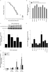
Figure 1. Quantitative expression of specific WNT pathway genes in human lung fibroblasts.
(A) WNT pathway gene expression in MRC5 human lung fibroblasts. Data shown are average Cq-values corrected for 18s ribosomal RNA expression determined in triplicate by quantitative real-time PCR. Of note; a lower Cq-value corresponds with higher gene expression. (B) The WNT pathway genes WNT-5A, WNT-5B, WNT-16, FZD2, FZD6, FZD8, DVL3 and β-catenin were analyzed by quantitative real-time PCR in MRC-5 fibroblasts and primary human lung fibroblasts. (C) Time-dependent activation of smad3 in response to TGF-β1 (2 ng/ml). Phosphorylation of ser423/425-smad3 was evaluated in whole cell lysates by immunoblotting using specific antibodies. Equal protein loading was verified by the analysis of total smad2/3. Data represents mean ± s.e.m. of 6 independent experiments. *p<0.05 compared to untreated MRC-5 fibroblasts determined by a two-tailed student's t-test for paired observations. (D) qRT-PCR analysis of WNT-5A, WNT-5B, WNT-16, FZD2, FZD6, FZD8, DVL3 and β-catenin in MRC-5 fibroblasts after 4 h of of TGF-β1 (2 ng/ml) stimulation. Expression of WNT pathway genes by TGF-β1 is corrected for 18S rRNA and expressed relative to untreated MRC-5 fibroblasts. Data represents mean ± s.e.m. of 5 independent experiments. *p<0.05, **p<0.01 compared to untreated MRC-5 fibroblasts determined by a two-tailed student's t-test for paired observations. (E) Effect of increasing concentrations TGF-β1 on WNT-5B, FZD8 and β-catenin gene expression. MRC-5 fibroblasts were stimulated with 0.5, 2.0 and 5.0 ng/ml TGF-β1 for 24 h. WNT-5B, FZD8 and β-catenin expression was determined by qRT-PCR analysis, corrected for 18S rRNA and expressed relative to untreated MRC-5 fibroblasts (control). Data represents mean ± s.e.m. of 4–7 independent experiments. p<0.05 for dose-dependency of WNT-5B, FZD8 and β-catenin gene expression in response to TGF-β1 determined by a One-way ANOVA.
Figure 2
Figure 2. Differential WNT pathway gene expression in primary lung fibroblasts from individuals with and without COPD.
Primary lung fibroblasts were isolated from individuals without (control) and with COPD (GOLD stage II and IV) as described in the materials en methods. qRT-PCR analysis of WNT-5A (A), WNT-5B (B), WNT-16 (C), FZD2 (D), FZD6 (E), FZD8 (F), DVL3 (G) and β-catenin (H) mRNA of primary lung fibroblasts treated with or without TGF-β1 (2 ng/ml) for 4 h. Expression of WNT pathway genes is plotted relative to the mean expression in untreated fibroblasts from controls. Data are derived from 7 controls and 11 COPD patients (5 GOLD stage II and 6 GOLD stage IV). mRNA expression was determined both at baseline (open circles; ○) and after TGF-β stimulation (closed circles; •). Median of each group is indicated by -----. *p<0.05, **p<0.01, ***p<0.001, two-tailed student's t-test for unpaired observations or a One-way ANOVA followed by a Newman-Keuls multiple comparison test.
Figure 3
Figure 3. TGF-β1 induces myofibroblast differentiation of MRC-5 lung fibroblasts.
MRC-5 fibroblasts were grown to confluence and treated for 24 h or 48 h with 2 ng/ml of TGF-β1. (A) Expression of the myofibroblasts markers α-sm-actin, fibronectin and MMP-2 was evaluated in whole cell lysates by immunoblotting using specific antibodies. Equal protein loading was verified by the analysis of GAPDH. Representative immunoblots of 5–8 independent experiments are shown. ***p<0.001, two-way student's t-test for paired observations. (B) Evaluation of stress fiber formation in MRC-5 lung fibroblasts after TGF-β1 stimulation. MRC-5 lung fibroblasts were treated for 48 h with TGF-β1 (2 ng/ml) and subsequently fixed and permeabilized. Cells were stained for filamentous actin (488 phalloidin; green) and nucleus (Hoechst 3342; blue). Pictures were taken at 400× magnification. (C) Effect of increasing concentrations TGF-β1 on myofibroblast differentiation. MRC-5 fibroblasts were stimulated with 0.5, 2.0 and 5.0 ng/ml TGF-β1 for 48 h. Expression of α-sm-actin, fibronectin and active β-catenin was evaluated in whole cell lysates by immunoblotting using specific antibodies. Equal protein loading was verified by the analysis of GAPDH. Representative immunoblots of 4 independent experiments are shown.
Figure 4
Figure 4. Treatment with TGF-β1 increases β-catenin signaling in MRC-5 lung fibroblasts.
MRC-5 fibroblasts were grown to confluence and treated for up to 24 h with TGF-β1 (2 ng/ml). (A) Expression of total β-catenin, active (non-phosphorylated) β-catenin and ser9/21 phosphorylation of GSK-3 were evaluated by immunoblotting using specific antibodies. Equal protein loading was verified by the analysis of GAPDH or total GSK-3, respectively. Responses of TGF-β1 on total and active β-catenin expression (B and C) and ser9/21-GSK-3 phosphorylation (D) were quantified by densitometry, representing mean ± s.e.m. of 3 independent experiments. *p<0.05, ***p<0.001, two-tailed student's t-test for paired observations or repeated measures ANOVA followed by a Newman-Keuls multiple comparison test. (E) Evaluation of cellular localization of active (non-phosphorylated) β-catenin in MRC-5 lung fibroblasts stimulated with TGF-β1 (2 ng/ml) for 48 h. Fixed and permeabilized MRC-5 fibroblasts were (immuno)cytochemically stained for active (non-phosphorylated) β-catenin (Cy3; red) and stained for filamentous actin (488 phalloidin; green) and nucleus (Hoechst 33342; blue). Pictures were taken at 400× magnification. (F) Increased cytosolic and nuclear expression of β-catenin in response to TGF-β1 stimulation. MRC-5 fibroblasts were stimulated with TGF-β1 (2 ng/ml) for 24 h. Subsequently cytosolic and nuclear extracts were prepared. Expression of total and active (non-phosphorylated) β-catenin was evaluated by immunoblotting. Equal protein loading was verified by the analysis of GAPDH and Lamin A/C, respectively. Representative immunoblots of 4 independent experiments are shown. (G) β-Catenin activation precedes myofibroblast differentiation. Expression of active β-catenin protein (open circles; ○), α-sm-actin mRNA (grey triangles; ▾) and α-sm-actin protein (black triangles; ▴) in response to TGF-β1 (2 ng/ml) was determined by immunoblotting and quantitative real time PCR. Data represents mean ± s.e.m. of 3–8 independent experiments.
Figure 5
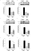
Figure 5. Silencing β-catenin expression by specific siRNA attenuates TGF-β1-induced α-sm-actin and fibronectin expression.
Subconfluent MRC-5 lung fibroblast cultures were transfected with a siRNA against the β-catenin transcript. Control cultures were transfected with a non-targeting control siRNA. Transfected cells were treated with TGF-β1 (2 ng/ml) for 48 h. (A–B) The efficiency of β-catenin silencing was evaluated by immunoblotting the expression of (A) total β-catenin and (B) active β-catenin and GAPDH to correct for differences in protein loading. Data represent mean ± s.e.m. of 4–6 experiments. *p<0.05, **p<0.01 and ***p<0.001 compared to non-targeting siRNA control, ###p<0.001 compared to non-targeting siRNA treated with TGF-β1 determined by a one-way ANOVA followed by a Newman-Keuls multiple comparison test. (C–F) β-catenin siRNA attenuated TGF-β1-induced α-sm-actin (C and E) and fibronectin (D and F) gene and protein expression. Expression or mRNA was determined by real-time PCR and normalized to 18S ribosomal mRNA expression. Protein expression was determined by immunoblotting and equal protein loading was verified by the analysis of GAPDH. Responses were quantified and normalized to the expression of 18S rRNA (gene) or GAPDH (protein). Data represent mean ± s.e.m. of 5–6 independent experiments. *p<0.05, ***p<0.001 compared to non-targeting siRNA control, #p<0.05, ##p<0.01 ###p<0.001 compared to non-targeting siRNA treated with TGF-β1, ††p<0.01 compared to β-catenin siRNA control, one-way ANOVA followed by a Newman-Keuls multiple comparison test.
Figure 6
Figure 6. Pharmacological inhibition of β-catenin attenuates TGF-β1-induced α-sm-actin and fibronectin expression.
Pharmacological inhibition of β-catenin/TCF4 signaling by quercetin or PKF115–584. Confluent MRC-5 lung fibroblasts were treated with TGF-β1 (2 ng/ml) for 48 h in the absence or presence of either quercetin (40 µM) or PKF115–584 (100 nM). Expression of α-sm-actin (A) and fibronectin (B) was evaluated by immunoblotting using a specific antibody. Responses were quantified by densitometry and normalized to the expression of GAPDH. Data represent mean ± s.e.m. of 3 independent experiments. ***p<0.001 compared untreated MRC-5 lung fibroblasts (control), ##p<0.01; ###p<0.001 to TGF-β1 treated MRC-5 lung fibroblasts determined by a one-way ANOVA followed by a Newman-Keuls multiple comparison test.
Figure 7
Figure 7. Increased β-catenin activation and fibronectin deposition in fibroblast of individuals with COPD in response to TGF-β1.
Primary lung fibroblasts were isolated from individuals without (control) and with COPD (GOLD stage II and IV) as described in the materials en methods. The fibroblasts were grown to confluence and treated for 48 h with TGF-β1 (2 ng/ml). Expression of active β-catenin (A) and fibronectin (B) was evaluated by immunoblotting. Equal protein loading was verified by the analysis of GAPDH. Responses were quantified by densitometry and normalized to the expression of GAPDH. Data are derived from 5 controls and 9 COPD patients (4 GOLD stage II and 5 GOLD stage IV). Median of each group is indicated by -----. *P<0.05. Statistical differences between control and COPD were determined a two-tailed Mann-whitney test.

References

    1. Pauwels RA, Buist AS, Ma P, Jenkins CR, Hurd SS. Global strategy for the diagnosis, management, and prevention of chronic obstructive pulmonary disease: National Heart, Lung, and Blood Institute and World Health Organization Global Initiative for Chronic Obstructive Lung Disease (GOLD): executive summary. Respir Care. 2001;46:798–825. - PubMed
    1. Rabe KF, Hurd S, Anzueto A, Barnes PJ, Buist SA, et al. Global strategy for the diagnosis, management, and prevention of chronic obstructive pulmonary disease: GOLD executive summary. Am J Respir Crit Care Med. 2007;176:532–555. - PubMed
    1. Barnes PJ, Shapiro SD, Pauwels RA. Chronic obstructive pulmonary disease: molecular and cellular mechanisms. Eur Respir J. 2003;22:672–688. - PubMed
    1. Konigshoff M, Kneidinger N, Eickelberg O. TGF-beta signaling in COPD: deciphering genetic and cellular susceptibilities for future therapeutic regimen. Swiss Med Wkly. 2009;139:554–563. - PubMed
    1. Chung KF. Cytokines in chronic obstructive pulmonary disease. Eur Respir J. 2001;(Suppl 34):50s–59s. - PubMed

Publication types

MeSH terms