. 2011;6(9):e25577.
doi: 10.1371/journal.pone.0025577. Epub 2011 Sep 28.

Human cysteine cathepsins are not reliable markers of infection by Pseudomonas aeruginosa in cystic fibrosis

Affiliations

Human cysteine cathepsins are not reliable markers of infection by Pseudomonas aeruginosa in cystic fibrosis

Clément Naudin et al. PLoS One. 2011.

Abstract

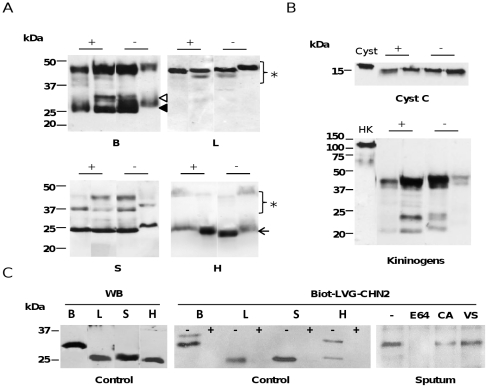
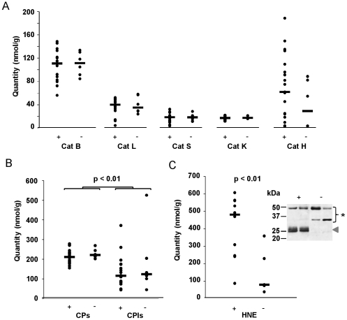
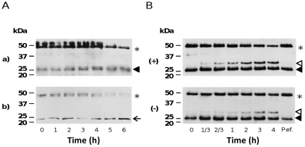
Cysteine cathepsins have emerged as new players in inflammatory lung disorders. Their activities are dramatically increased in the sputum of cystic fibrosis (CF) patients, suggesting that they are involved in the pathophysiology of CF. We have characterized the cathepsins in CF expectorations and evaluated their use as markers of colonization by Pseudomonas aeruginosa. The concentrations of active cathepsins B, H, K, L and S were the same in P. aeruginosa-positive (19 Ps+) and P. aeruginosa-negative (6 Ps-) samples, unlike those of human neutrophil elastase. Also the cathepsin inhibitory potential and the cathepsins/cathepsin inhibitors imbalance remained unchanged and similar (∼2-fold) in the Ps+ and Ps- groups (p<0.001), which correlated with the breakdown of their circulating cystatin-like inhibitors (kininogens). Procathepsins, which may be activated autocatalytically, are a potential proteolytic reservoir. Immunoblotting and active-site labeling identified the double-chain cathepsin B, the major cathepsin in CF sputum, as the main molecular form in both Ps+ and Ps- samples, despite the possible release of the ∼31 kDa single-chain form from procathepsin B by sputum elastase. Thus, the hydrolytic activity of cysteine cathepsins was not correlated with bacterial colonization, indicating that cathepsins, unlike human neutrophil elastase, are not suitable markers of P. aeruginosa infection.


Conflict of interest statement

Competing Interests: The authors have declared that no competing interests exist.

Figures

Figure 1
Figure 1. Cysteine cathepsins and their inhibitors in supernatants of CF sputum.
Only representative samples are shown. (A) Proteins (30 µg/well) were separated by 15% SDS-PAGE under reducing conditions, transferred to nitrocellulose membranes, and analyzed with polyclonal antibodies against human cathepsins B, H, L and S. (+): Pseudomonas aeruginosa-colonized CF sputum; (−): Pseudomonas aeruginosa-negative CF sputum.◃, single-chain cathepsin B; ◂, double-chain cathepsin B; ←, mature cathepsins S and H; ★, proforms. (B) Immunostaining with polyclonal anti-cystatin C antibody and anti-kininogen antibody. (+): Pseudomonas aeruginosa-colonized CF sputum; (−): Pseudomonas aeruginosa-negative CF sputum. Control: Cyst, Cystatin C; HK, HMWK. Recombinant human cystatin C (R&D systems) has an additional C-terminal 10 His-tag and an apparent molecular mass of 17 kDa, according to the supplier. (C) Supernatants of CF sputum incubated with Biot-LVG-CHN2 (30 µM), for 1 h at 37°C . Other samples were pre-incubated with E-64, CA-074, and Mu-Leu-Hph-VSPh prior to adding the biotinylated activity-based probe. Samples were separated by 12% SDS-PAGE, electroblotted and incubated with extravidin-peroxidase conjugate. The peroxidase activity was revealed by chemiluminescence. WB: individual cathepsins B, H, L and S immunoblotted as control. Control: (−), no pre-incubation with E-64; (+), pre-incubation with E-64 prior to adding Biot-LVG-CHN2. Sputum: E-64, pre-incubation with E-64; CA, pre-incubation with CA-074; VS, pre-incubation with Mu-Leu-HphVSPh.
Figure 2
Figure 2. Degradation of human HMWK by CF sputum.
Exogenous HMWK was incubated in the activity buffer with supernatants of CF sputum at 30°C for 0–5 hours. Hydrolysis products were separated by 12.5% SDS-PAGE, transferred to nitrocellulose membranes and immunoblotted with rabbit polyclonal anti-kininogen antibody . Lane 1, 0-h incubation; lane 2, 2-h incubation; lane 3, 5-h incubation; lane 4, 5-h incubation in the presence of CA-074; lane 5, 5-h incubation in the presence of E-64. For clarity, one representative sample is shown.
Figure 3
Figure 3. CP and HNE activities, and CP/CPI balance in CF sputum.
(+): P. aeruginosa-colonized CF sputum; (−): P. aeruginosa-negative CF sputum. Cathepsins B, H, K, L and S and HNE activities were quantified as reported in details in the experimental section. Data are shown as individual points and statistically significant P values are shown. The horizontal bars indicate medians. (A) Cathepsins B, L, S, K and H. (B) CP/CPI balance. CPI: expressed as inhibitory site (cystatin-like) equivalent. (C) Elastase activities: the horizontal bars indicate medians. The western blot analysis was performed using a rabbit anti-HNE antibody (representative samples are shown). ◂, unbound HNE; ★, bound HNE.
Figure 4
Figure 4. Maturation of human procathepsins in CF sputum.
(A) Autocatalytic maturation of procathepsins B and S. Supernatants were incubated for up to 6 h in 100 mM sodium acetate buffer pH 4.3, 10 µg/ml dextran sulfate, 4 mM DTT, at 37°C. The maturation products were separated by 12% SDS/PAGE under reducing conditions, transferred to a nitrocellulose membrane and analyzed by western blotting using polyclonal antibodies specific for (a) human cathepsin B and (b) human cathepsin S. One representative sample is shown. ★, proforms; ◂, mature double-chain cathepsin B; ←, mature cathepsin S. (B) Elastase-dependent maturation of procathepsin B. Supernatants were incubated for up to 4 h in 50 mM HEPES pH 7.4, 150 mM NaCl, 0.05% NP40 at 37°C and the maturation products analyzed by immunoblotting using a polyclonal anti-cathepsin B antibody. Pefabloc was used as control (4 h). Pef, Pefabloc (AEBSF). (+): P. aeruginosa-positive CF sputum (one representative sample); (−): P. aeruginosa-negative CF sputum (one representative sample). ◃, single-chain cathepsin B; ◂, double-chain cathepsin B; ★, procathepsin B.

References

    1. Kerem B, Rommens JM, Buchanan JA, Markiewicz D, Cox TK, et al. Identification of the cystic fibrosis gene: genetic analysis. Science. 1989;245:1073–1080. - PubMed
    1. Riordan JR, Rommens JM, Kerem B, Alon N, Rozmahel R, et al. Identification of the cystic fibrosis gene: cloning and characterization of complementary DNA. Science. 1989;245:1066–1073. - PubMed
    1. Grasemann H, Ratjen F. Emerging therapies for cystic fibrosis lung disease. Expert Opin Emerg Drugs. 2010;15:653–659. - PubMed
    1. Pham CT. Neutrophil serine proteases: specific regulators of inflammation. Nat Rev Immunol. 2006;6:541–550. - PubMed
    1. Korkmaz B, Moreau T, Gauthier F. Neutrophil elastase, proteinase 3 and cathepsin G: physicochemical properties, activity and physiopathological functions. Biochimie. 2008;90:227–242. - PubMed

Publication types