. 2011;6(9):e25633.
doi: 10.1371/journal.pone.0025633. Epub 2011 Sep 28.

Excitotoxicity triggered by Neurobasal culture medium

Affiliations

Excitotoxicity triggered by Neurobasal culture medium

Joshua Hogins et al. PLoS One. 2011.

Abstract

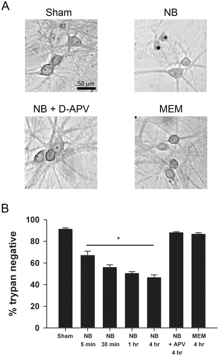
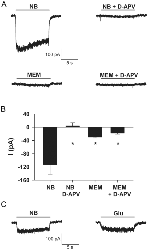
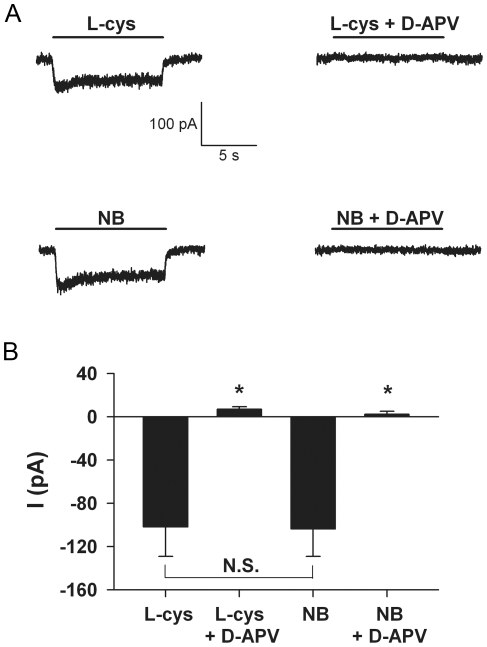
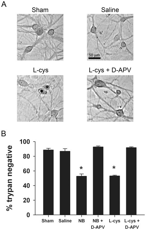
Neurobasal defined culture medium has been optimized for survival of rat embryonic hippocampal neurons and is now widely used for many types of primary neuronal cell culture. Therefore, we were surprised that routine medium exchange with serum- and supplement-free Neurobasal killed as many as 50% of postnatal hippocampal neurons after a 4 h exposure at day in vitro 12-15. Minimal Essential Medium (MEM), in contrast, produced no significant toxicity. Detectable Neurobasal-induced neuronal death occurred with as little as 5 min exposure, measured 24 h later. D-2-Amino-5-phosphonovalerate (D-APV) completely prevented Neurobasal toxicity, implicating direct or indirect N-methyl-D-aspartate (NMDA) receptor-mediated neuronal excitotoxicity. Whole-cell recordings revealed that Neurobasal but not MEM directly activated D-APV-sensitive currents similar in amplitude to those gated by 1 µM glutamate. We hypothesized that L-cysteine likely mediates the excitotoxic effects of Neurobasal incubation. Although the original published formulation of Neurobasal contained only 10 µM L-cysteine, commercial recipes contain 260 µM, a concentration in the range reported to activate NMDA receptors. Consistent with our hypothesis, 260 µM L-cysteine in bicarbonate-buffered saline gated NMDA receptor currents and produced toxicity equivalent to Neurobasal. Although NMDA receptor-mediated depolarization and Ca²⁺ influx may support survival of young neurons, NMDA receptor agonist effects on development and survival should be considered when employing Neurobasal culture medium.


Conflict of interest statement

Competing Interests: The authors have declared that no competing interests exist.

Figures

Figure 1
Figure 1. Fresh Neurobasal is neurotoxic to cultured hippocampal neurons.
A. Photomicrographs from a single experiment showing toxicity resulting from fresh Neurobasal (NB) incubation. Upper left: brightfield image from a control sham medium exchange stained with trypan blue 24 h after the challenge. Upper right: a field from a dish incubated in fresh Neurobasal for 4 h. Pyknotic, trypan positive nuclei with remnants of cell bodies are present. Lower left: 50 µM D-APV, a competitive NMDA receptor antagonist, was included in the fresh Neurobasal medium and protected neurons from Neurobasal toxicity. Lower right: A field from a dish incubated for 4 h in MEM, matched to Neurobasal for inorganic salts, osmolarity, pH, glucose, and glycine concentrations. B. Summary of experiments like that of panel A showing the effects of Neurobasal (NB) exposure over various incubation times. MEM incubation had minimal effect (N = 4 independent platings, asterisk indicates a main effect of Neurobasal compared with sham condition; p<0.01, one-way ANOVA).
Figure 2
Figure 2. Acute Neurobasal application (diluted 50% in recording saline) generates a D-APV-sensitive current in synaptically isolated hippocampal neurons grown in microculture.
A. Representative currents from a cell demonstrating the response to 50% Neurobasal (NB) in the absence and presence of 50 µM D-APV. MEM application (50% dilution) produced minimal current. In this and subsequent figures, cells were voltage-clamped at −30 mV to relieve Mg2+ block of the NMDA receptor channel. Scale bar applies to all traces. B. Summary of the steady-state current amplitudes from 5 cells. Asterisk designates p<0.05 compared with sham (unpaired, two-tailed t-tests with Bonferroni correction for multiple comparisons). C. Representative traces from a cell demonstrating similar current amplitude generated by acutely applied Neurobasal (NB) diluted to 50% in standard saline and 0.5 µM glutamate (Glu) added to BBS. The scale bar applies to both traces. The faster onset kinetics and offset kinetics of Neurobasal-gated currents compared with glutamate-gated currents were a consistent finding (see Results). These calibrations suggest that undiluted Neurobasal contains a low-affinity agonist, equivalent to 1 µM glutamate.
Figure 3
Figure 3. L-cysteine, a component of Neurobasal, elicits a D-APV-sensitive current similar to Neurobasal.
A. Representative currents from a hippocampal neuron showing the response to acute application of 130 µM L-cysteine (L-cys) and 50% Neurobasal (NB). The current was blocked by co-application of 50 µM D-APV. Acute application of L-cysteine in BBS and Neurobasal generated similar D-APV-sensitive current amplitude and kinetics. The calibration bar applies to all traces. B. Summary of steady-state current amplitude from 7 cells. Asterisk designates p<0.05 compared with current amplitude in the absence of D-APV (unpaired, two-tailed t-tests with Bonferroni correction for multiple comparisons). N.S. designates no statistical significance (p>0.05 using paired, two-tailed t tests).
Figure 4
Figure 4. L-cysteine and Neurobasal generate similar toxicity.
A. Photomicrographs from a single experiment showing toxicity as a result of L-cysteine (260 µM) incubation. Upper left: brightfield image from a sham medium exchange control. Upper right: A field from a dish incubated for 4 h in BBS (saline). Lower left: a field from a dish exposed to BBS plus 260 µM L-cysteine for 4 h. Trypan-positive nuclei with remnants of cell bodies are present. Lower right: 50 µM D-APV protected against L-cysteine-induced toxicity. B. Summary of experiments like that of panel A compared with Neurobasal (NB) incubation in sibling cultures. Neurotoxicity of 260 µM L-cysteine in BBS is similar to Neurobasal toxicity, and both are blocked by co-incubation with 50 µM D-APV (N = 4 independent experiments on independent platings). Asterisk designates p<0.05 compared with sham (unpaired, two-tailed t-tests with Bonferroni correction for multiple comparisons).

References

    1. Brewer GJ, Torricelli JR, Evege EK, Price PJ. Optimized survival of hippocampal neurons in B27-supplemented Neurobasal, a new serum-free medium combination. J Neurosci Res. 1993;35:567–576. - PubMed
    1. Olney JW, Zorumski C, Price MT, Labruyere J. L-cysteine, a bicarbonate-sensitive endogenous excitotoxin. Science. 1990;248:596–599. - PubMed
    1. Shute AA, Cormier RJ, Moulder KL, Benz A, Isenberg KE, et al. Astrocytes exert a pro-apoptotic effect on neurons in postnatal hippocampal cultures. Neuroscience. 2005;131:349–358. - PubMed
    1. Choi DW, Rothman SM. The role of glutamate neurotoxicity in hypoxic-ischemic neuronal death. Annu Rev Neurosci. 1990;13:171–182. - PubMed
    1. Rothman SM. The neurotoxicity of excitatory amino acids is produced by passive chloride influx. J Neurosci. 1985;5:1483–1489. - PMC - PubMed

Publication types