. 2011;6(9):e25639.
doi: 10.1371/journal.pone.0025639. Epub 2011 Sep 27.

Dopamine-induced plasticity, phospholipase D (PLD) activity and cocaine-cue behavior depend on PLD-linked metabotropic glutamate receptors in amygdala

Affiliations

Dopamine-induced plasticity, phospholipase D (PLD) activity and cocaine-cue behavior depend on PLD-linked metabotropic glutamate receptors in amygdala

Balaji Krishnan et al. PLoS One. 2011.

Abstract

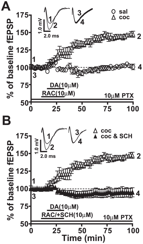
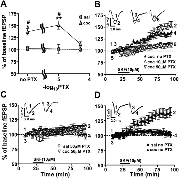
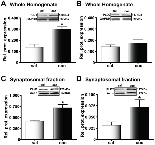
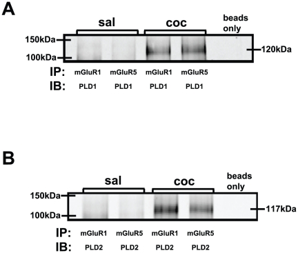
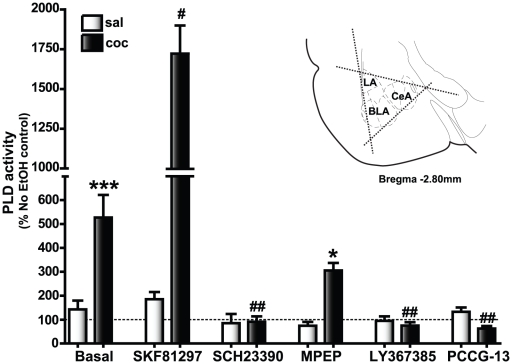
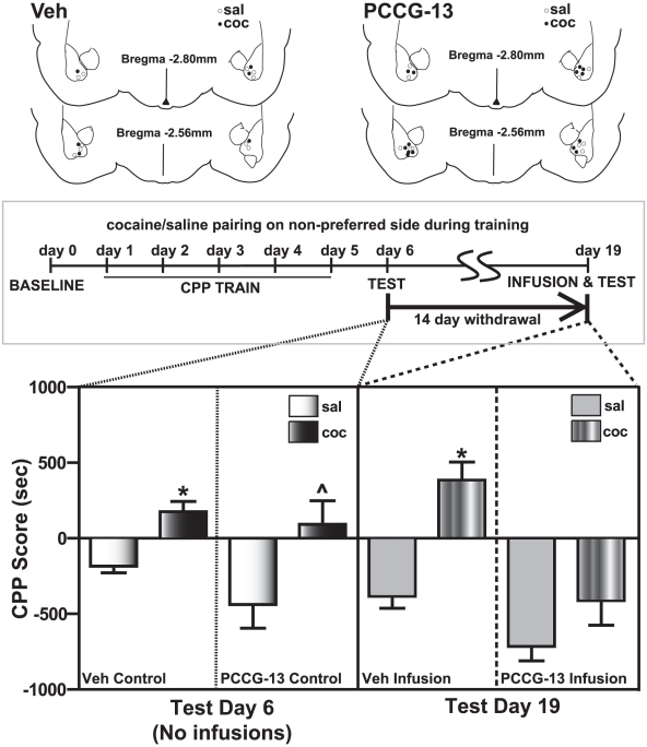
Cocaine-cue associations induce synaptic plasticity with long lasting molecular and cellular changes in the amygdala, a site crucial for cue-associated memory mechanisms. The underlying neuroadaptations can include marked alterations in signaling via dopamine (DA) receptors (DRs) and metabotropic glutamate (Glu) receptors (mGluRs). Previously, we reported that DR antagonists blocked forms of synaptic plasticity in amygdala slices of Sprague-Dawley rats withdrawn from repeated cocaine administration. In the present study, we investigated synaptic plasticity induced by exogenous DA and its dependence on mGluR signaling and a potential role for phospholipase D (PLD) as a downstream element linked to mGluR and DR signaling. Utilizing a modified conditioned place preference (CPP) paradigm as a functional behavioral measure, we studied the neurophysiological effects after two-weeks to the last cocaine conditioning. We recorded, electrophysiologically, a DR-induced synaptic potentiation in the basolateral to lateral capsula central amygdala (BLA-lcCeA) synaptic pathway that was blocked by antagonists of group I mGluRs, particularly, the PLD-linked mGluR. In addition, we observed 2-2.5 fold increase in PLD expression and 3.7-fold increase in basal PLD enzyme activity. The enhanced PLD activity could be further stimulated (9.3 fold) by a DA D1-like (D1/5R) receptor agonist, and decreased to control levels by mGluR1 and PLD-linked mGluR antagonists. Diminished CPP was observed by infusion of a PLD-linked mGluR antagonist, PCCG-13, in the amygdala 15 minutes prior to testing, two weeks after the last cocaine injection. These results imply a functional interaction between D1/5Rs, group I mGluRs via PLD in the amygdala synaptic plasticity associated with cocaine-cues.


Conflict of interest statement

Competing Interests: The authors have declared that no competing interests exist.

Figures

Figure 1
Figure 1. Amygdala slices (B) from animals exhibiting robust cocaine CPP (A) measured 14 days after the last day of CPP training demonstrate a D1/5R agonist-induced LTP in the BLA-lcCeA pathway (C) which is abolished by D1/5R antagonist application (D).
A) The cocaine CPP group (black bars) had significantly greater CPP scores than saline-treated animals (white bars) irrespective of whether the drug pairing was on the preferred (saline: 187.1±75.1, cocaine: 448.2±55.7, *p<0.05, n = 34) or the non-preferred side (saline: −239.7±78.5, cocaine: 203.8±71.7, ***p<0.005, n = 34). B) Placement of recording (Rec) and stimulating (Stim) electrodes are indicated in a schematic representation of the slice containing BLA-lcCeA pathway using a rat brain atlas template . C) D1/5R agonist (SKF81297) induces LTP in the BLA-lcCeA pathway in brain slices from animals conditioned to cocaine and tested two weeks after the last CPP training day (clear triangles, 151.4±8.8%, *p<0.05, n = 6). The saline-treated group did not show potentiation (clear circles, 101.6±9.7%, ns, n = 7). Responses are plotted as percent change from the baseline field EPSPs as a function of time. Numbers on the representative traces show the time on the graph at which they were recorded. D) SKF81297-induced LTP in the amygdala from slices of cocaine CPP animals (clear triangles) is completely abolished by the D1/5R antagonist, SCH23390 (filled triangles, 94.5±10.9%, **p<0.01, n = 4). Significance is denoted by increasing number of asterisks (*). For comparison panels C and D use same data graphs and fEPSP traces for the slices from cocaine CPP group superfused with SKF81297.
Figure 2
Figure 2. The endogenous neurotransmitter, dopamine (DA), in the presence of raclopride (RAC), a D2R antagonist, induced LTP mediated through D1/5R activation.
Responses are plotted as percent change of baseline fEPSPs as a function of time. Numbers on the representative traces show the time on the graph at which they were recorded. A) DA in the presence of RAC (clear triangles, 146.5±3.2%, *p<0.05, n = 5) shows potentiation similar to that recorded with SKF81297 (Figure 1B) in amygdala slices from cocaine CPP animals while the saline-treated group (clear circles, 102.2±2.4%, ns, n = 5) exhibits no LTP. B) DA+RAC-induced LTP is abolished by SCH23390 in the cocaine CPP group (filled triangles, 94.5±4.5%, ns, n = 6). For comparison panels A and B use same data graphs and fEPSP traces for the slices from cocaine CPP group superfused with SKF81297.
Figure 3
Figure 3. GABAergic inhibition in the BLA-lcCeA synapse is essential for SKF81297-induced LTP in the cocaine CPP group.
Field EPSP magnitude is plotted with respect to baseline values as a function of increasing PTX concentrations. Responses are plotted as percent change from the baseline fEPSPs as a function of time. Numbers on the representative traces show the time on the graph at which they were recorded. A) In the cocaine CPP group (filled triangles), SKF81297-induced LTP is lost when GABAergic inhibition is blocked with 50 µM PTX (saline: 101.1±5.1%, cocaine: 112.2±4.6%, ns, n = 7). In the saline-treated groups (filled circles: 102.6±4.0%, 101.6±9.7% and 101.1±5.1%, ns, n = 5–7), the SKF81297-associated fEPSPs do not show a dependence on the extent of GABAergic inhibition. B) SKF81297-induced LTP at different concentrations of PTX in amygdala slices from the cocaine CPP group is plotted as a function of time. LTP in 50 µM PTX (inverted triangles, 112.2±4.6%, n = 7) is inhibited compared to LTP in 10 µM PTX (clear triangles, 151.4±8.8%, n = 6) or LTP in no PTX (filled triangles, 137.5±6.9%, n = 5). C) SKF81297-induced LTP in 50 µM PTX (inverted clear triangles) in the cocaine CPP group is diminished to levels recorded in the saline-treated group (filled circles). D) With GABAergic inhibition intact, SKF81297-induced LTP is significantly increased in the cocaine CPP group (filled triangles) compared to the saline-treated group (filled circles). Panels use same data graphs as in A, B and fEPSP traces in B to illustrate the comparisons. For comparison, all panels use same data graphs and fEPSP traces for the slices from cocaine CPP group superfused with SKF81297 in 10 µM PTX represented in Figures 1 and 2.
Figure 4
Figure 4. SKF81297-induced LTP in the cocaine CPP group is dependent on the PLD-linked mGluR, mGluR1, and partially dependent on mGluR5 and PLC activity.
Responses are plotted as percent change from the baseline fEPSPs as a function of time. Numbers on the representative traces show the time on the graph at which they were recorded. A) SKF81297-induced LTP in the cocaine CPP group (clear triangles, 151.4±8.8%, *p<0.05, n = 6) is completely blocked by the PLD-linked mGluR antagonist (PCCG-13, filled triangles, 95.0±9.2%, n = 6). B) mGluR1 receptor antagonist (LY367385, filled triangles, 106.0±6.7%, n = 6) blocks the SKF81297-induced LTP (clear triangles, *p<0.05, n = 6). C) The mGluR5 antagonist (MPEP, filled triangles, 122.7±5.6%, n = 6) significantly reduces but does not abolish the SKF81297-induced LTP (clear triangles, *p<0.05, n = 6). D) PLC antagonist (U-73122, filled triangles, 128.2±6.1%, n = 6), reduces but does not eliminate the SKF81297-induced LTP (clear triangles, *p<0.05, n = 6). For comparison, panels A and B use same data graphs and fEPSP traces for the slices from cocaine CPP group superfused with SKF81297 as shown in Figures 1, 2 and 3.
Figure 5
Figure 5. PLD levels in the amygdala are increased in cocaine CPP animals.
Protein expression relative to the loading control is plotted along the Y-axis. Representative immunoblots are shown in the panels above each graph; *p<0.05 compared to the corresponding saline-treated control. A) PLD1 levels in the whole amygdala homogenate are significantly increased in cocaine CPP animals (black bars) compared to the saline-treated group (white bars). B) PLD2 levels in the whole amygdala homogenate are not increased in the cocaine CPP group (black bars) compared to the saline-treated group (white bars). C) In the amygdala crude synaptosomal fraction, PLD1 protein levels are increased in the cocaine CPP group (black bars) suggesting that there is increased synaptosomal membrane incorporation of PLD1 in this experimental group compared to the saline-treated group (white bars). D) Similar to PLD1, amygdala crude synaptosomal levels of PLD2 show an increase in the cocaine CPP group (black bars). However, such increased expression is observed despite a lack of increase in the whole homogenate levels, suggesting that recruitment from the existing pool of PLD2 to the synaptosomal membrane is increased in the cocaine CPP group.
Figure 6
Figure 6. Amygdala protein subjected to co-immunoprecipitation (IP) with mGluR1, mGluR5 and subsequently immunoblotted (IB) with PLD antibodies show association between PLD1/2 and mGluRs1/5 in the cocaine CPP group.
A) PLD1 and B) PLD2 were detected only in the amygdala of cocaine CPP group (coc) but not saline-treated group (sal).
Figure 7
Figure 7. Basal PLD activity is strongly stimulated by the D1/5R agonist and blocked by the D1/5R, mGluR5, mGluR1, and the PLD-linked mGluR antagonists in the amygdala of cocaine CPP animals.
The dotted line indicates PLD activity associated with control slices (no EtOH added) which was determined for each animal and used to calculate the change in PLD activity levels with EtOH and/or drug application. Basal levels represent the increase in PLD activity observed in the EtOH-treated slices compared to the no EtOH controls; *p<0.05 compared to the corresponding saline control and # p<0.05 compared to the cocaine CPP group basal PLD activity. Basal PLD activity was significantly increased (***p<0.001, n = 50) in the cocaine CPP group (dark bars, 527.3±94.3) compared to the saline-treated group (white bars, 142.6±36.9). SKF81297, the D1/5R agonist, application increased the basal levels in the cocaine CPP group significantly (1722.0±176.9, n = 12, # p<0.05) compared to the basal PLD activity observed with EtOH treatment alone in the same experimental group. The D1/5R antagonist, SCH23390, completely blocked basal PLD activity (91.2±21.9, n = 12, ## p<0.01) in the cocaine CPP group. A similar reduction in PEtOH levels was observed with application of either the PLD-linked mGluR antagonist, PCCG-13 (62.9±10.6, n = 7, ## p<0.01) or the mGluR1 antagonist, LY367385 (75.0±13.9, n = 12, ## p<0.01), while the mGluR5 antagonist, MPEP, did not decrease basal PLD activity (305.7±31.5, n = 7, ns) within the cocaine CPP group but were significantly increased compared to (*p<0.05) the saline treated group. Applications of SKF81297 (184.9±30.5, n = 12), SCH23390 (84.9±38.9, n = 12), MPEP (74.2±16.3, n = 7), LY367385 (94.7±18.9, n = 12) and PCCG-13 (132.5±18.4, n = 7) did not significantly alter the PEtOH levels in the saline-treated group compared to the basal activity levels. Inset is a depiction of the triangular excision performed to isolate amygdala (bilaterally for each animal, each slice) containing the basolateral (BLA), the central (CeA) and the lateral (LA) subregions from three serial coronal slices (350 µm) beginning −2.30 mm to −2.80 mm from Bregma .
Figure 8
Figure 8. Expression of cocaine CPP was blocked by bilateral infusions of the PLD-linked mGluR antagonist into the amygdala of cocaine CPP group 14 days after the last CPP training.
Infusions of the vehicle into the cocaine CPP animals (striped bars, 383.5±121.0, n = 3, Veh infusion panel) on test day 19 do not diminish cocaine CPP (black bars, 176.1±56.3, n = 3, Veh control panel) measured on test day 6. No significant changes were observed in the behavioral response of the saline-treated group between day 6 (white bars, −170.2±36.2, n = 5, Veh control panel) and day 19 (grey bars, −369.2±71.3, n = 5, Veh infusion panel), suggesting that the vehicle infusions alone had no effect. PCCG-13 infusion 15 minutes prior to CPP testing results in a marked loss of preference for the drug-associated side (black bars, cocaine day 6: 92.4±145.9, striped bars, cocaine day 19: −397.1±155.6, *p<0.05, n = 7, PCCG-13 control and PCCG-13 infusion panels respectively) in the cocaine CPP animals, while no significant change in the behavioral response is observed in saline-treated group (white bars, saline day 6: −423.5±149.7, grey bars, saline day 19: −700.8±143.2, ns, n = 5, PCCG-13 control and PCCG-13 infusion panels respectively). *p<0.05 compared to the corresponding vehicle control (saline) and by ∧p<0.05 compared to the corresponding vehicle control (PCCG-13). Cannulae placement for the animals is depicted (above the graph) for both the vehicle (Veh) and drug (PCCG-13). These sites were mapped to BLA on rat brain atlas templates. A schematic representation of the experimental protocol utilized for CPP training along with the two days of CPP testing is illustrated above the corresponding data in the graph below.

Similar articles

Cited by

References

    1. Hyman SE. Addiction: a disease of learning and memory. Am J Psychiatry. 2005;162:1414–1422. - PubMed
    1. Buffalari DM, See RE. Amygdala mechanisms of Pavlovian psychostimulant conditioning and relapse. Curr Top Behav Neurosci. 2010;3:73–99. - PubMed
    1. Hyman SE, Malenka RC, Nestler EJ. Neural mechanisms of addiction: the role of reward-related learning and memory. Annu Rev Neurosci. 2006;29:565–598. - PubMed
    1. Koob GF, Volkow ND. Neurocircuitry of addiction. Neuropsychopharmacology. 2010;35:217–238. - PMC - PubMed
    1. Breiter HC, Gollub RL, Weisskoff RM, Kennedy DN, Makris N, et al. Acute effects of cocaine on human brain activity and emotion. Neuron. 1997;19:591–611. - PubMed

Publication types

MeSH terms