. 2011 Sep;5(9):e1331.
doi: 10.1371/journal.pntd.0001331. Epub 2011 Sep 27.

Scabies mite peritrophins are potential targets of human host innate immunity

Affiliations

Scabies mite peritrophins are potential targets of human host innate immunity

Angela Mika et al. PLoS Negl Trop Dis. 2011 Sep.

Erratum in

Abstract

Background: Pruritic scabies lesions caused by Sarcoptes scabiei burrowing in the stratum corneum of human skin facilitate opportunistic bacterial infections. Emerging resistance to current therapeutics emphasizes the need to identify novel targets for protective intervention. We have characterized several protein families located in the mite gut as crucial factors for host-parasite interactions. Among these multiple proteins inhibit human complement, presumably to avoid complement-mediated damage of gut epithelial cells. Peritrophins are major components of the peritrophic matrix often found in the gut of arthropods. We hypothesized that a peritrophin, if abundant in the scabies mite gut, could be an activator of complement.

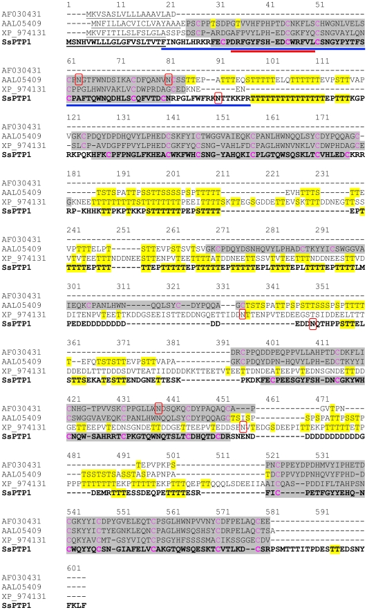
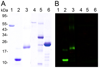
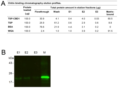
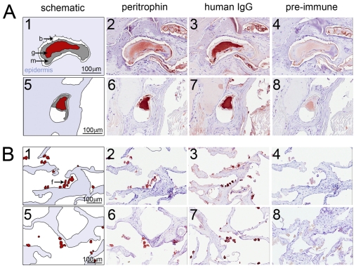
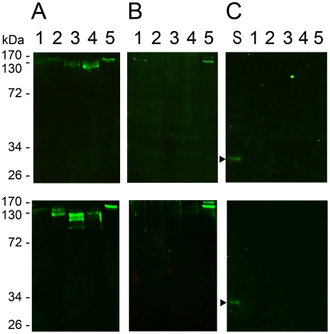
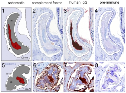
Methodology/principal findings: A novel full length scabies mite peritrophin (SsPTP1) was identified in a cDNA library from scabies mites. The amino acid sequence revealed four putative chitin binding domains (CBD). Recombinant expression of one CBD of the highly repetitive SsPTP1 sequence as TSP-hexaHis-fusion protein resulted in soluble protein, which demonstrated chitin binding activity in affinity chromatography assays. Antibodies against a recombinant SsPTP1 fragment were used to immunohistochemically localize native SsPTP1 in the mite gut and in fecal pellets within the upper epidermis, co-localizing with serum components such as host IgG and complement. Enzymatic deglycosylation confirmed strong N- and O-glycosylation of the native peritrophin. Serum incubation followed by immunoblotting with a monoclonal antibody against mannan binding lectin (MBL), the recognition molecule of the lectin pathway of human complement activation, indicated that MBL may specifically bind to glycosylated SsPTP1.

Conclusions/significance: This study adds a new aspect to the accumulating evidence that complement plays a major role in scabies mite biology. It identifies a novel peritrophin localized in the mite gut as a potential target of the lectin pathway of the complement cascade. These initial findings indicate a novel role of scabies mite peritrophins in triggering a host innate immune response within the mite gut.


Conflict of interest statement

The authors have declared that no competing interests exist.

Figures

Figure 1
Figure 1. Full length amino acid sequence of scabies mite peritrophin 1 (SsPTP1) aligned with other arthropod peritrophins.
The scabies mite peritrophin SsPTP1 is shown in bold in a sequence alignment with Anopheles gambiae peritrophin 1 (AF030431), peritrophin from Aedes agypti (AAL05409) and a peritrophin matrix protein from Tribolium castaneum (XP_974131). The following predictions are shown: signal sequences (underlined); chitin binding domains (grey background); N-glycosylated residues (red box); O-glycosylated residues (yellow background). Cysteine residues predicted to form disulfide bonds within the chitin binding domains are shown in magenta. A blue and a red bar indicate the sequence of the chitin binding domain CBD1 and the peptide c2, respectively.
Figure 2
Figure 2. Specificity of mouse antisera raised against a recombinant chitin binding domain of SsPTP1.
Shown is a series of purified scabies mite proteins on a coomassie-stained SDS-PAGE (A) and a corresponding western with a peritrophin-specific antibody (B). The gel (A) demonstrates the purity of the recombinant peptide CBD1 (lane 2) and the fusion protein TSP-CBD1 (lane 3) and (B) the specificity of the antibody raised against CBD1, confirming no cross-reaction with unrelated scabies mite proteins SMSB3a (lane 1), SMIPP-S D1 (lane 5), SMIPP-S I1 (lane 6) and BSA (lane 4).
Figure 3
Figure 3. Binding of recombinant peritrophin fusion protein TSP-CBD1 by affinity chromatography to chitin magnetic beads.
(A) Shown are elution profiles of the peritrophin fusion protein TSP-CBD1, Schistosoma mansoni TSP protein and bovine serum albumin (BSA) (negative controls), and wheat germ agglutinin (WGA; positive control). Data are means of n = 4 independent experiments measured in duplicate. 100 µg protein was loaded in each chromatography run. E1, elution (8M urea); E2, elution (5% SDS); E3, elution (10% SDS); matrix-bound, the amount of protein estimated to be still bound to the matrix after E1-E3 elution; n.d., not detectable (B) Western blotting of chitin binding chromatography fractions from an representative TSP-CBD1 run. Immunodetection was performed using polyclonal mouse antibodies against peritrophin peptide CBD1. Lanes E1-E3 show elution fractions and lane M protein bound to chitin matrix beads.
Figure 4
Figure 4. Immunohistological localisation of SsPTP1 in scabies mite gut and excreted mite feces.
Histological sections were probed with antibodies raised against the peritrophin peptide CBD1 (A2, B2), against peptide c2 (A6, B6), with anti-human IgG (A3, A7, B3, B7) and corresponding pre-immune sera (A4, A8, B4, B8). Red staining indicates binding of primary antibody. A schematic diagram in the left panel (A1 and A5; B1 and B5) outlines the features in the histological sections. The mite gut and fecal pellets are shown in red, the mite body in gray, the burrow in white and the epidermis in blue. b, burrow; f, feces; g, gut; m, mite. Scale bars (100 µM) indicate the magnification level.
Figure 5
Figure 5. Deglycosylation of native scabies mite peritrophin 1 and binding to the human complement factor MBL.
Scabies mite homogenates were incubated for 24 hours (upper panel) or 3 days (bottom panel) in either buffer alone (lane 1), or with PNGase F (lane 2), four O-glycosidases (lane 3), or PNGase F and four O-glycosidases (lane 4) at 37°C. The preparations were separated by SDS-PAGE in comparison with a sample of fresh mite homogenate (lane 5). Western blots were probed for SsPTP1 using anti-CBD1 sera (A), or for MBL using an anti-MBL antibody after incubation of the blot with 50% normal human serum (B) or with buffer without serum (C). In blot (C) an additional lane was loaded with 1% normal human serum in PBS (lane S). Arrows point to a single band corresponding to the 32kDa subunits of human MBL. The experiment was repeated three times.
Figure 6
Figure 6. Immunohistological localisation detects complement factor C9, but not the activated SC5b-9 complex in scabies mite infested human skin sections.
Histological sections were probed with antibodies raised against human SC5b-9 neoantigen (2) and against complement factor C9 (6), with anti-human IgG (3 and 7) and the respective pre-immune sera from goat (4) and rabbit (8). Red staining indicates binding of primary antibody. A schematic diagram in the left panel (1 and 5) outlines the features in the histological sections. The mite gut and fecal pellets are shown in red, the mite body in gray, the burrow in white and the epidermis in blue. b, burrow; f, feces; g, gut; m, mite. Scale bars (100 µM) indicate the magnification level.

Similar articles

Cited by

References

    1. Hengge UR, Currie BJ, Jäger G, Lupi O, Schwartz RA. Scabies: a ubiquitous neglected skin disease. The Lancet Infectious Diseases. 2006;6:769–779. - PubMed
    1. Arlian LG. Biology, host relations and epidemiology of Sarcoptes scabiei. Annual Review of Entomology. 1989;34:139–161. - PubMed
    1. Mellanby K. The development of symptoms, parasitic infection and immunity in human scabies. Parasitology. 1944;35:197–206.
    1. Walton SF, Holt DC, Currie BJ, Kemp DJ, Baker, et al. Advances in Parasitology: Academic Press; 2004. Scabies: New Future for a Neglected Disease. pp. 309–376. - PubMed
    1. Currie BJ, Carapetis JR. Skin infections and infestations in Aboriginal communities in northern Australia. Australas J Dermatol. 2000;41:139–143; quiz 144–135. - PubMed

Publication types

MeSH terms

Associated data