. 2012 Jan;13(1):43-53.
doi: 10.1111/j.1600-0854.2011.01299.x. Epub 2011 Nov 8.

Post-Golgi supramolecular assembly of aquaporin-4 in orthogonal arrays

Affiliations

Post-Golgi supramolecular assembly of aquaporin-4 in orthogonal arrays

Andrea Rossi et al. Traffic. 2012 Jan.

Abstract

The supramolecular assembly of aquaporin-4 (AQP4) in orthogonal arrays of particles (OAPs) involves N-terminus interactions of the M23-AQP4 isoform. We found AQP4 OAPs in cell plasma membranes but not in endoplasmic reticulum (ER) or Golgi, as shown by: (i) native gel electrophoresis of brain and AQP4-transfected cells, (ii) photobleaching recovery of green fluorescent protein-AQP4 chimeras in live cells and (iii) freeze-fracture electron microscopy (FFEM). We found that AQP4 OAP formation in plasma membranes, but not in the Golgi, was not related to AQP4 density, pH, membrane lipid composition, C-terminal PDZ domain interactions or α-syntrophin expression. Remarkably, however, fusion of AQP4-containing Golgi vesicles with (AQP4-free) plasma membrane vesicles produced OAPs, suggesting the involvement of plasma membrane factor(s) in AQP4 OAP formation. In investigating additional possible determinants of OAP assembly we discovered membrane curvature-dependent OAP assembly, in which OAPs were disrupted by extrusion of plasma membrane vesicles to ∼110 nm diameter, but not to ∼220 nm diameter. We conclude that AQP4 supramolecular assembly in OAPs is a post-Golgi phenomenon involving plasma membrane-specific factor(s). Post-Golgi and membrane curvature-dependent OAP assembly may be important for vesicle transport of AQP4 in the secretory pathway and AQP4-facilitated astrocyte migration, and suggests a novel therapeutic approach for neuromyelitis optica.


Figures

Figure 1
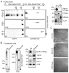
Figure 1. AQP4 oligomerization in subcellular fractions of brain homogenates and transfected cells
(A) Electrophoresis of human brain plasma membrane, Golgi and ER fractions. The first dimension is BN/PAGE, the second dimension is tricine SDS/PAGE. Immunodetection was carried out against AQP4 or CX43 (left). Immunoblot of Na+/K+ ATPase, TGN46 and calnexin (right). (B) BN/PAGE of plasma membrane and Golgi/ER fractions of U87MG cells transfected with M23-AQP4 (left) or CX43 (center). Immunoblot of Na+/K+ ATPase, TGN46 and calnexin (right). (C) Unidirectionally-shadowed freeze-fracture electron micrographs of purified subcellular fractions of M1 and M23-AQP4-transfected U87MG cells. Scale bar, 200 nm.
Figure 2
Figure 2. Oligomerization of AQP4-GFP chimeras and localization in live cells
(A) SDS/PAGE (top left), BN/PAGE (top center) and hrCN/PAGE (top right) of M1-GFP and M23-GFP. Confocal and TIRF microscopy showing plasma membrane targeting of M1-GFP and M23-GFP in transiently transfected U87MG cells (bottom). (B) Live-cell imaging of U87MG cells transiently transfected with M1-GFP and M23-GFP after low-temperature block showing Golgi localization (top), or treatment with BFA showing ER localization (bottom). Cells were co-transfected with compartment-specific marker proteins (GalT for Golgi, calnexin for ER) fused to mCherry.
Figure 3
Figure 3. Diffusion of M1 and M23-AQP4 in live cells
(A) FRAP of M1-GFP or M23-GFP in transiently transfected U87MG cells, with plasma membrane (top) and Golgi (low-temperature treated cells, bottom) localization. Arrowheads indicate bleached area. (B) Representative kinetics of FRAP of M1-GFP and M23-GFP over 120 s (top), with summary of percentage recovery data at 120 s (bottom) (S.E., n > 10 cells, * P < 0.01).
Figure 4
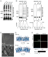
Figure 4. Membrane curvature-dependent AQP4 oligomerization
(A) BN/PAGE of M23-AQP4 in transfected U87MG cells after incubation in buffers of indicated pH. (B) (left) BN/PAGE of M23-AQP4 in plasma membrane vesicles of different sizes generated by extrusion (top). Vesicle size distributions determined by quasi-elastic light scattering (bottom). (right) BN/PAGE of M23-AQP4 in control and extruded plasma membrane vesicles, and in extruded vesicles after fusion. (C) Freeze-fracture electron micrographs of vesicles of average diameters 355 nm (top) and 57 nm (bottom). (D) To-scale model of AQP4 tetramers in membranes of different curvatures correspond to vesicles of diameters 350 or 50 nm. (E) Fluorescence micrographs of coverslip-immobilized 355 and 57 nm diameter vesicles showing binding of NMO monoclonal antibody rAb-53 (red) and anti-AQP4 antibody (green). Red-to-green fluorescence ratios (R/G) (right) (mean ± S.E., n=3).
Figure 5
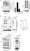
Figure 5. Determinants of AQP4 oligomerization on the plasma membrane
(A) Effect of cholesterol depletion on M23-AQP4 oligomerization in plasma membrane vesicles from M23-AQP4-expressing U87MG cells. (left) BN/PAGE after treatment with indicated concentration of βMCD. (center) BN/PAGE after treatment with indicated concentrations of SMase to deplete SM (S.E., n=4). (right) Cholesterol content in Golgi and plasma membrane vesicles treated with βMCD (S.E., n=4). (B) BN/PAGE of M23-AQP4 oligomerization in unfused and high Ca2+-fused mixtures of plasma membrane and Golgi vesicles. As indicated, vesicles were derived from M23-AQP4-expressing or non-transfected cells. (right) Vesicle size distribution before (pre-fusion) and after incubation in high Ca2+ buffer. (C) BN/PAGE of AQP4 after α-syntrophin siRNA knock-down in human astrocyte cultures (bottom). Knock-down (α-syn) and loading (actin) controls are shown (top). (D) BN/PAGE of U87MG cells after transfection with native M23-AQP4 or a C-terminus truncation mutant lacking the PDZ-binding domain (bottom). SDS/PAGE of α-syntrophin expression is shown (top).

References

    1. Frigeri A, Gropper MA, Turck CW, Verkman AS. Immunolocalization of the mercurial-insensitive water channel and glycerol intrinsic protein in epithelial cell plasma membranes. Proc Natl Acad Sci U S A. 1995;92:4328–4331. - PMC - PubMed
    1. Frigeri A, Gropper MA, Umenishi F, Kawashima M, Brown D, Verkman AS. Localization of MIWC and GLIP water channel homologs in neuromuscular, epithelial and glandular tissues. J Cell Sci. 1995;108 (Pt 9):2993–3002. - PubMed
    1. Ho JD, Yeh R, Sandstrom A, Chorny I, Harries WE, Robbins RA, Miercke LJ, Stroud RM. Crystal structure of human aquaporin 4 at 1. 8 A and its mechanism of conductance. Proc Natl Acad Sci U S A. 2009;106:7437–7442. - PMC - PubMed
    1. Verkman AS, Binder DK, Bloch O, Auguste K, Papadopoulos MC. Three distinct roles of aquaporin-4 in brain function revealed by knockout mice. Biochim Biophys Acta. 2006;1758:1085–1093. - PubMed
    1. Lennon VA, Kryzer TJ, Pittock SJ, Verkman AS, Hinson SR. IgG marker of optic-spinal multiple sclerosis binds to the aquaporin-4 water channel. J Exp Med. 2005;202:473–477. - PMC - PubMed

Publication types

MeSH terms

LinkOut - more resources