. 2012 Jan;14(1):133-46.
doi: 10.1111/j.1462-5822.2011.01708.x. Epub 2011 Nov 10.

The immunity-related GTPase Irgm3 relieves endoplasmic reticulum stress response during coxsackievirus B3 infection via a PI3K/Akt dependent pathway

Affiliations

The immunity-related GTPase Irgm3 relieves endoplasmic reticulum stress response during coxsackievirus B3 infection via a PI3K/Akt dependent pathway

Zhen Liu et al. Cell Microbiol. 2012 Jan.

Abstract

The IRG protein Irgm3 preserves cell survival during coxsackievirus B3 (CVB3) infection. However, the molecular mechanisms are not clear. Here, we examined the effect of Irgm3 expression on ER stress triggered by pharmacological agents or CVB3 infection. In Tet-On/Irgm3 HeLa cells, Irgm3 expression suppressed either chemical- or CVB3-induced upregulation of glucose-regulated protein 78. Further, Irgm3 strongly inhibited the activation of both the PERK and ATF6 pathways of ER stress responses, which further led to the diminished phosphorylation of eIF2α, reduced cleavage/activation of transcription factor SREBP1 and attenuated induction of proapoptotic genes CHOP and GADD34. These data were further supported by experiments using Irgm3 knockout mouse embryonic fibroblasts, in which the ER stress induced by CVB3 was not relieved due to the lack of Irgm3 expression. In addition, the tunicamycin-triggered ER stress promoted the subsequent CVB3 infection. The effect of Irgm3 on ER stress and CVB3 infection was diminished by the PI3K inhibitor, LY294002, while inhibitors of ERK, JNK and p38 had no effect. These data were further corroborated by transfection of cells with a dominant negative Akt. Taken together, these data suggest that Irgm3 relieves the ER stress response via a PI3K/Akt dependent mechanism, which contributes to host defence against CVB3 infection.


Figures

Figure 1
Figure 1. Irgm3 relieves ER stress response triggered by chemical inducers
Tet-On/Irgm3 HeLa cells were induced with 1 μg/ml Dox, and Western blot analysis was conducted to confirm that Dox itself has no effect on GRP78 expression in normal condition (A). Tet-On/Irgm3 HeLa cells were induced with 1 μg/ml Dox for 16 h and then treated with Tu (B) or BFA (C) at the indicated concentrations. Cell lysates were harvested at 16 h and 20 h post treatment with Tu and BFA respectively, and then subjected to immunoblotting using indicated antibodies. (D). Premade Tet-On HeLa cells were transfected with pTRE vector, pTRE-Irgm3/S98N mutant or pTRE-WT Irgm3, and induced with Dox for 36 hrs. Then, cells were treated with 1 μg/ml Tunicamycin and cell lysates were collected for immunoblotting. All Data were quantitated by densitometric analysis and normalized to the β-actin expression. Untreated controls were arbitrarily set to 1.0, and expression of GRP78 was calculated as fold increase as compared to the control group. Data represents the mean ± S.E. from three independent experiments (*p<0.05).
Figure 2
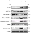
Figure 2. Irgm3 inhibits the activation of PERK pathway of ER stress response
Tet-On/Irgm3 HeLa cells were induced with 1 μg/ml Dox for 16 h and then treated with 1 μg/ml Tu for 16 h or untreated. Cell lysates were subjected to immunoblot analysis with indicated antibodies. Detection of β-actin was used as the loading control.
Figure 3
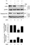
Figure 3. Irgm3 inhibits the activation of ATF6 pathway of ER stress response
Tet-On/Irgm3 HeLa cells were induced with Dox and then treated with Tu or untreated as described in Fig. 2. Cell fractionation was performed, and cytosolic and nuclear fractions were collected separately. Proform and cleaved forms of ATF6 were detected in the cytosolic and nuclear fraction, respectively. Results were quantitated by densitometric analysis and normalized to the β-actin and histone-1 expression, respectively. Untreated controls were arbitrarily set to 1.0, and levels of ATF6 were calculated as fold increase compared to the control group. Data represents the mean ± S.E. from three independent experiments (*p<0.05).
Figure 4
Figure 4. Irgm3 expression inhibits the ER stress-mediated apoptotic response
Tet-On/Irgm3 HeLa cells were induced with 1 μg/ml Dox for 16 h and then treated with 1 μg/ml Tu for 20 h or untreated. Cell lysates were harvested and subjected to immunoblot analysis with indicated antibodies. β-actin was used as the loading control.
Figure 5
Figure 5. Irgm3 inhibits the CVB3-induced upregulation of GRP78
(A). Tet-On/Irgm3 HeLa cells were treated with 1 μg/ml Dox for 20 h or untreated and then infected with CVB3 at 10 MOI or sham infected with PBS. Cell lysates were collected at the indicated time points pi and subjected to immunoblotting. GRP78 expression was quantitated by densitometric analysis and normalized to the β-actin expression. The value of GRP78 expression for the uninduced sample at 0 h was arbitrarily set to 1.0, and fold changes of other samples were calculated as compared to the control group. Data represents the mean ± S.E. from three independent experiments (*p<0.05). (B). To confirm that vector or Dox itself has no effect on GRP78 expression, premade Tet-On HeLa cells were induced with Dox and then infected with CVB3 or sham-infected as described above. Cell lysates were collected at the indicated time points pi and subjected to immunoblotting for GRP78 and Irgm3 expression.
Figure 6
Figure 6. Irgm3 inhibits the activation of PERK and ATF6 pathways during CVB3 infection
(A). Tet-On/Irgm3 HeLa cells were treated with 1 μg/ml Dox for 20 h or untreated and then infected with CVB3 at 10 MOI. Cell lysates were collected at the indicated time points pi and subject to immunoblot analysis using indicated antibodies. The expression of p-eIF2 α were quantitated by densitometric analysis and normalized to the total eIF2 α expression. The value of p-eIF2 α for the uninduced control sample at 0 h was arbitrarily set to 1.0, and fold change of other samples were calculated based on the value of control. The data represents the mean ± S.E. from three independent experiments (*p<0.05). (B). Cells were induced with Dox for 20 h and then infected with CVB3 at 10 MOI or sham-infected with PBS for 12 h. Cell fractions were prepared, and the proform and cleaved forms of ATF6 were detected in the nuclear and cytosolic fraction, respectively. Histone-1 and β-actin expression levels were used as loading controls.
Figure 7
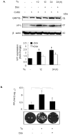
Figure 7. Preemptive ER stress response induced by Tu facilitates viral protein synthesis and viral particle release
Dox-induced or uninduced Tet-On/Irgm3 HeLa cells at 16 h post induction were treated with or without 1μg/ml Tu for indicated hours and then infected with CVB3 at 10 MOI for 10 h. Cell lysates were collected and subjected to immunoblot analysis using indicated antibodies. CVB3 VP1 protein expression was quantitated by densitometric analysis and normalized to the β-actin expression. VP1 level of untreated cells was arbitrarily set to 1.0, and fold increase of other samples was calculated based on the untreated control. Data represents the mean ± S.E. from three independent experiments (*p<0.05) (A). Dox induced or uninduced Tet-On/Irgm3 HeLa cells were treated with or without 1μg/ml Tu for 12 h and then infected with CVB3 for 10 h at 10 MOI. Supernatants only (B) and supernatants plus cells (C) from the cell cultures were collected respectively, and the pfu/ml was determined by plaque assays on HeLa cell monolayers. Data represents the mean ± S.E from three independent experiments (*p<0.05).
Figure 8
Figure 8. Irgm3 relieves CVB3-induced ER stress response in mouse embryonic fibroblasts (MEF)
WT MEF and Irgm3 KO MEF were treated with 500 U/ml IFN- γ for indicated hours. Cell lysates were collected after treatment and Irgm3 expression was probed by immunoblotting (A). WT and KO MEF were infected with CVB3 at 40 MOI or sham-infected with PBS in the presence or absence of 500U/ml IFN- γ. Cell lysates were harvested at 30 h pi and subjected to immunoblot analysis using indicated antibodies. GRP78 and VP1 expression were quantitated by densitometric analysis and normalized to the β-actin expression respectively. Untreated/sham-infected control from either cell line was arbitrarily set to 1.0, and fold increase of other samples were calculated based on the value of their respective controls. Data represents the mean ± S.E from three independent experiments (*p<0.05) (B). Supernatants only (C) and supernatants plus cells (D) from the cell cultures that were treated as described in (B) were collected respectively, and the pfu/ml was determined by plaque assays on HeLa cell monolayers. Data represents the mean ± S.E from three independent experiments (*p<0.05).
Figure 9
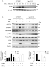
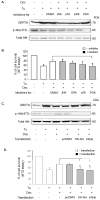
Figure 9. The relief of ER stress response by Irgm3 depends on the activation of PI3K/Akt pathway
(A). Dox-induced or uninduced Tet-On/Irgm3 HeLa cells were treated either with vehicle (DMSO) or with specific inhibitors including PI3K inhibitor LY294002 at 10 μM, ERK inhibitor U0126 at 20 μM_p38 inhibitor SB203580 at 20 μM, and JNK inhibitor SP600125 at 40 μM. Cells were then treated with 1 μg/ml Tu for 16 h. Cell lysates were harvested and subjected to immunoblot analysis. Total Akt was used as a loading control. (B). After cells were treated with inhibitors and Tu as described above, cell viability was measured by MTS assay. Absorbance values were corrected by subtracting the background reading. The value of MTS assay in untreated cells was defined as 100% survival (control). The cell viability of other samples was expressed relative to the control. Data represents the mean ± S.E from three independent experiments (*p<0.05). (C). Tet-On/Irgm3 HeLa cells were transiently transfected with a plasmid vector, or a plasmid encoding DN-Akt or FRNK using Lipofectamine 2000. At 24 h post transfection, cells were then treated with 1 μg/ml Tu for 16 h and then subjected to immunoblot analysis. (D). Cells treated under the same conditions as described in (C) were also subjected to MTS assay. The value of cell viability of untreated cells was defined as 100% survival (control). The cell viability of other samples was expressed relative to the control. Data represents the mean ± S.E from three independent experiments (*p<0.05).

References

    1. Alwine JC. Modulation of host cell stress responses by human cytomegalovirus. Curr Top Microbiol Immunol. 2008;325:263–279. - PubMed
    1. Averous J, Bruhat A, Jousse C, Carraro V, Thiel G, Fafournoux P. Induction of CHOP expression by amino acid limitation requires both ATF4 expression and ATF2 phosphorylation. J Biol Chem. 2004;279:5288–5297. - PubMed
    1. Baltzis D, Qu LK, Papadopoulou S, Blais JD, Bell JC, Sonenberg N, Koromilas AE. Resistance to vesicular stomatitis virus infection requires a functional cross talk between the eukaryotic translation initiation factor 2alpha kinases PERK and PKR. J Virol. 2004;78:12747–12761. - PMC - PubMed
    1. Bekpen C, Hunn JP, Rohde C, Parvanova I, Guethlein L, Dunn DM, et al. The interferon-inducible p47 (IRG) GTPases in vertebrates: loss of the cell autonomous resistance mechanism in the human lineage. Genome Biol. 2005;6:R92. - PMC - PubMed
    1. Bienz K, Egger D, Troxler M, Pasamontes L. Structural organization of poliovirus RNA replication is mediated by viral proteins of the P2 genomic region. J Virol. 1990;64:1156–1163. - PMC - PubMed

Publication types

MeSH terms

Substances