Review
. 2011 Oct 7;44(1):9-16.
doi: 10.1016/j.molcel.2011.09.003.

RIPK-dependent necrosis and its regulation by caspases: a mystery in five acts

Affiliations
Review

RIPK-dependent necrosis and its regulation by caspases: a mystery in five acts

Douglas R Green et al. Mol Cell. .

Abstract

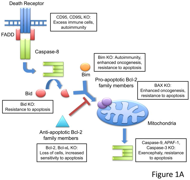
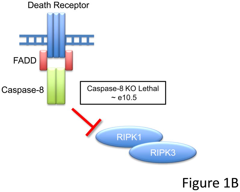
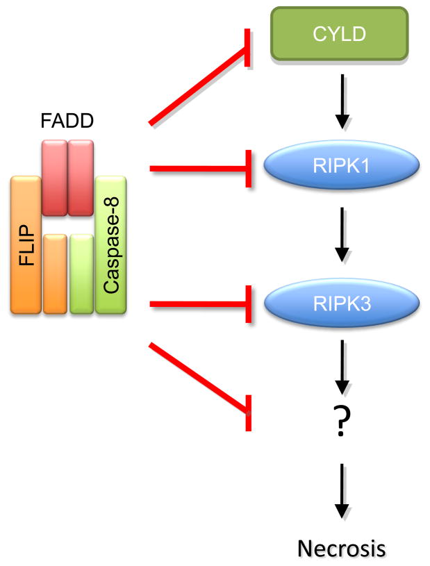
Caspase-8, FADD, and FLIP orchestrate apoptosis in response to death receptor ligation. Mysteriously however, these proteins are also required for normal embryonic development and immune cell proliferation, an observation that has led to their implication in several nonapoptotic processes. While many scenarios have been proposed, recent genetic and biochemical evidence points to unregulated signaling by the receptor-interacting protein kinases-1 (RIPK1) and RIPK3 as the lethal defect in caspase-8-, FADD-, and FLIP-deficient animals and tissues. The RIPKs are known killers, being responsible for a nonapoptotic form of cell death with features similar to necrosis. However, the mechanism by which caspase-8, FADD, and FLIP prevent runaway RIPK activation is unknown, and the signals that trigger these events during development and immune cell activation remain at large. In this review, we will lay out the evidence as it now stands, reinterpreting earlier observations in light of new clues and considering where the investigation might lead.


Figures

Figure 1
Figure 1
(A) Schematic of apoptotic death pathways. Extrinsic signals through death receptors like CD95 or intrinsic signals that activate pro-apoptotic Bcl-2 members like Bid and Bim lead to activation of downstream effector caspases (caspase-3). Loss of pro-apoptotic molecules lead to a range of phenotypes including embryonic lethality associated with exencephaly (caspase-9, APAF-1, or caspase-3 knockouts), and lymphoaccumulative and autoimmune disorders (CD95/CD95L or Bim knockout mice). (It should be noted, however, that while cells lacking downstream elements of the mitochondrial pathway do not undergo apoptosis via this route, cells may nevertheless die by a caspase-independent mechanism). Additionally, animals deficient in certain pro-apoptotic molecules (i.e. Bim or Bax knockouts) display enhanced oncogenesis. Cells from animals where pro-apoptotic proteins are deleted are generally resistant to apoptosis, while cells from animals missing anti-apoptotic proteins (such as Bcl-2 and Bcl-xL) have increased sensitivity to apoptotic stimuli. (B) Ablation of Caspase-8 leads to embryonic lethality circa e10.5. The simultaneous deletion of Caspase-8 and RIPK3 or FADD together with RIPK1 rescue embryonic development, suggesting that FADD and Caspase-8 act in concert to provide survival signals which may inhibit RIPK function during development.
Figure 1
Figure 1
(A) Schematic of apoptotic death pathways. Extrinsic signals through death receptors like CD95 or intrinsic signals that activate pro-apoptotic Bcl-2 members like Bid and Bim lead to activation of downstream effector caspases (caspase-3). Loss of pro-apoptotic molecules lead to a range of phenotypes including embryonic lethality associated with exencephaly (caspase-9, APAF-1, or caspase-3 knockouts), and lymphoaccumulative and autoimmune disorders (CD95/CD95L or Bim knockout mice). (It should be noted, however, that while cells lacking downstream elements of the mitochondrial pathway do not undergo apoptosis via this route, cells may nevertheless die by a caspase-independent mechanism). Additionally, animals deficient in certain pro-apoptotic molecules (i.e. Bim or Bax knockouts) display enhanced oncogenesis. Cells from animals where pro-apoptotic proteins are deleted are generally resistant to apoptosis, while cells from animals missing anti-apoptotic proteins (such as Bcl-2 and Bcl-xL) have increased sensitivity to apoptotic stimuli. (B) Ablation of Caspase-8 leads to embryonic lethality circa e10.5. The simultaneous deletion of Caspase-8 and RIPK3 or FADD together with RIPK1 rescue embryonic development, suggesting that FADD and Caspase-8 act in concert to provide survival signals which may inhibit RIPK function during development.
Figure 2
Figure 2
(A) Caspase-8 consists of a prodomain, followed by the large and the small protease subunits, separated by linker regions. The prodomain has two death “folds” named Death Effector Domains (DEDs, light blue), through which caspase-8 is recruited and dimerized. Upon dimerization, Caspase-8 become proteolytically active and processes itself in specific cleavage sites present in the linker regions. The cleavage between the large and small subunit contributes to the stabilization of the molecule and is essential to induction of cell death but is dispensable for RIPK-necrosis inhibition. (B) FLIP has a similar structure than Caspase-8, but lacks the catalytic cysteine. Although it can act as a dominant negative for caspase-8-induced death when overexpressed, FLIP acts in concert with Caspase-8 to inhibit RIPK-induced necrosis.
Figure 3
Figure 3
Caspase-8-FLIP complex blocks RIPK-dependent necrosis, although the precise mechanism behind this inhibition is still unknown. RIPK1 and RIPK3, as well as upstream deubiquitinase CYLD, are caspase-8 cleavage targets, suggesting that cleavage of any of these proteins might be critical for necrosis inhibition. However, as little is known about what is downstream RIPK1/RIPK3 activation, other targets for caspase-8 may yet be implicated.

References

    1. Alappat EC, Feig C, Boyerinas B, Volkland J, Samuels M, Murmann AE, Thorburn A, Kidd VJ, Slaughter CA, Osborn SL, et al. Phosphorylation of FADD at serine 194 by CKIalpha regulates its nonapoptotic activities. Mol Cell. 2005;19:321–332. - PubMed
    1. Baines CP, Kaiser RA, Purcell NH, Blair NS, Osinska H, Hambleton MA, Brunskill EW, Sayen MR, Gottlieb RA, Dorn GW, et al. Loss of cyclophilin D reveals a critical role for mitochondrial permeability transition in cell death. Nature. 2005;434:658–662. - PubMed
    1. Barbero S, Barila D, Mielgo A, Stagni V, Clair K, Stupack D. Identification of a critical tyrosine residue in caspase 8 that promotes cell migration. J Biol Chem. 2008;283:13031–13034. - PMC - PubMed
    1. Barbero S, Mielgo A, Torres V, Teitz T, Shields DJ, Mikolon D, Bogyo M, Barila D, Lahti JM, Schlaepfer D, et al. Caspase-8 association with the focal adhesion complex promotes tumor cell migration and metastasis. Cancer Res. 2009;69:3755–3763. - PMC - PubMed
    1. Bertrand MJ, Milutinovic S, Dickson KM, Ho WC, Boudreault A, Durkin J, Gillard JW, Jaquith JB, Morris SJ, Barker PA. cIAP1 and cIAP2 facilitate cancer cell survival by functioning as E3 ligases that promote RIP1 ubiquitination. Mol Cell. 2008;30:689–700. - PubMed

MeSH terms

LinkOut - more resources