. 2011 Oct 6;72(1):57-71.
doi: 10.1016/j.neuron.2011.08.033.

Exogenous α-synuclein fibrils induce Lewy body pathology leading to synaptic dysfunction and neuron death

Affiliations

Exogenous α-synuclein fibrils induce Lewy body pathology leading to synaptic dysfunction and neuron death

Laura A Volpicelli-Daley et al. Neuron. .

Abstract

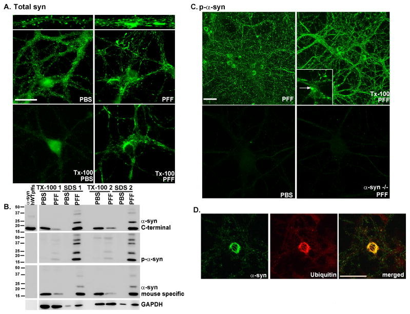
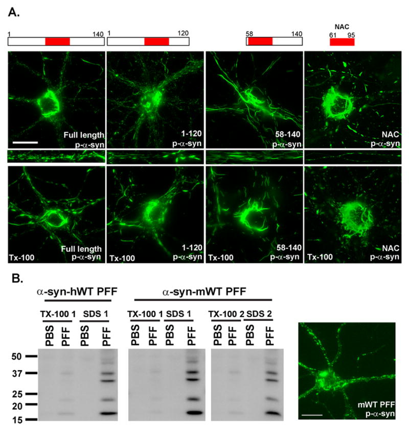
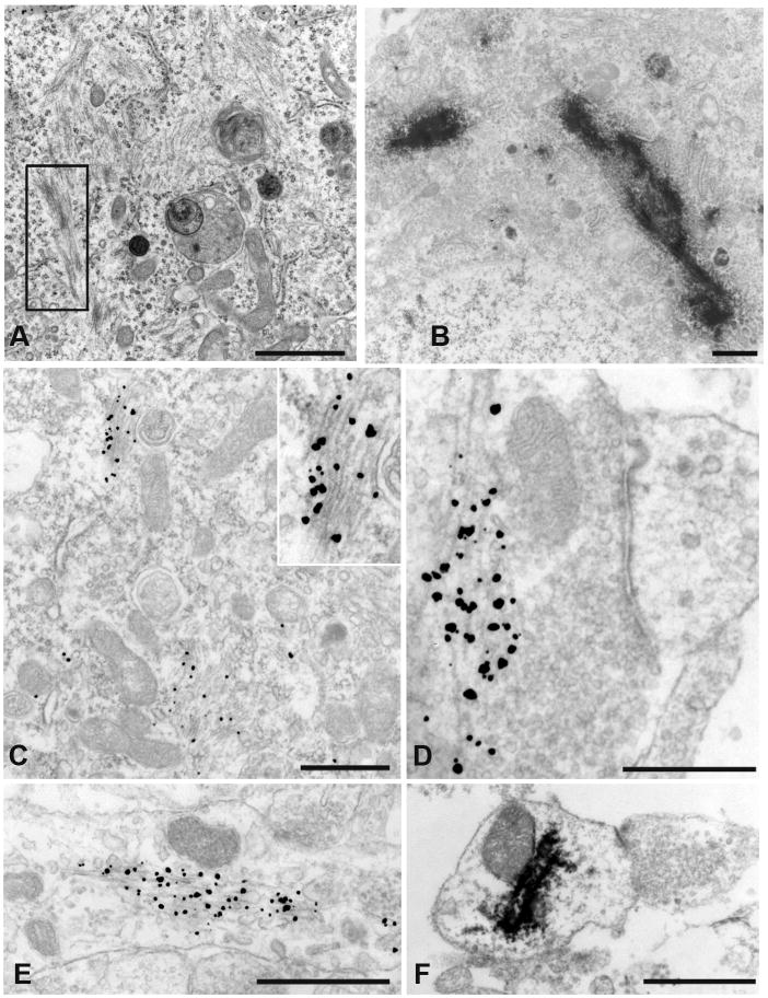
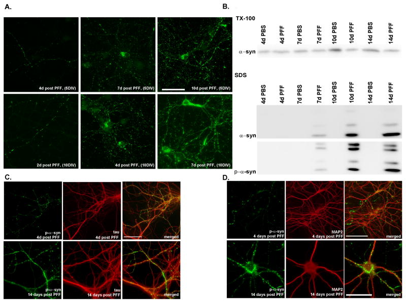
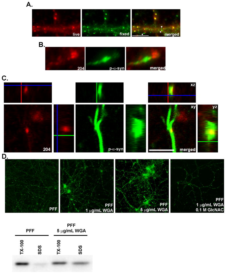
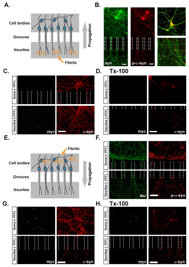
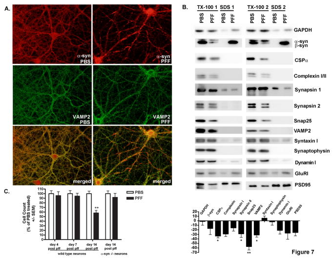
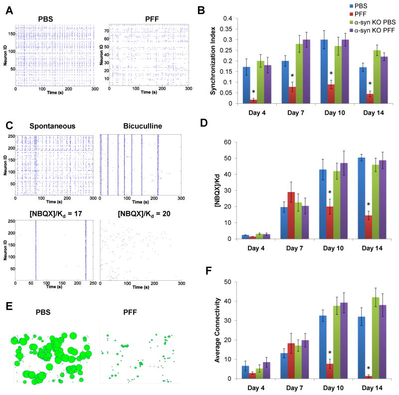
Inclusions composed of α-synuclein (α-syn), i.e., Lewy bodies (LBs) and Lewy neurites (LNs), define synucleinopathies including Parkinson's disease (PD) and dementia with Lewy bodies (DLB). Here, we demonstrate that preformed fibrils generated from full-length and truncated recombinant α-syn enter primary neurons, probably by adsorptive-mediated endocytosis, and promote recruitment of soluble endogenous α-syn into insoluble PD-like LBs and LNs. Remarkably, endogenous α-syn was sufficient for formation of these aggregates, and overexpression of wild-type or mutant α-syn was not required. LN-like pathology first developed in axons and propagated to form LB-like inclusions in perikarya. Accumulation of pathologic α-syn led to selective decreases in synaptic proteins, progressive impairments in neuronal excitability and connectivity, and, eventually, neuron death. Thus, our data contribute important insights into the etiology and pathogenesis of PD-like α-syn inclusions and their impact on neuronal functions, and they provide a model for discovering therapeutics targeting pathologic α-syn-mediated neurodegeneration.


Figures

Figure 1
Figure 1. α-syn-hWT pffs recruit endogenous α-syn to form pathologic, insoluble aggregates
A. Two weeks following pff addition, neurons were fixed with paraformaldehyde alone or paraformaldehyde with 1% Tx-100 to extract soluble proteins. In PBS treated neurons, α-syn localized to the presynaptic terminal and was Tx-100 soluble. Addition of α-syn pffs formed Tx-100 insoluble aggregates which recruited α-syn away from synapses. Scale bar = 20 μm. B. Neurons were treated with α-syn-hWT pffs, and 2 weeks later, were sequentially extracted with 1% Tx-100 followed by 2% SDS. Shown are immunoblots from 2 independent sets of samples. Antibodies that either recognize the C-terminus of α-syn (top) or are specific for mouse α-syn (bottom) showed that in PBS treated neurons, α-syn was soluble in Tx-100. α-Syn-hWT pff treatment reduced soluble α-syn and increased Tx-100-insoluble α-syn. The first lane shows α-syn-hWT pffs alone to demonstrate that the C-terminal antibody recognizes both human and mouse α-syn, and the mouse specific antibody recognizes only mouse pffs. Furthermore, the α-syn-hWT pffs themselves are not phosphorylated. C. Addition of α-syn pffs increased pathologic, p-α-syn. Fixation with paraformaldehyde/Tx-100 demonstrated that the phosphorylated aggregates were insoluble. Scale bar = 50 μm. Insert: Within the somata, aggregates appear as LB-like skein-like filaments and dense inclusions (arrow). PBS- treated neurons and neurons from α-syn −/− mice did not show p-α-syn. D. The phosphorylated aggregates are ubiquitin positive (N=2). Scale bar = 50 μm. See also Supplementary Figure 1.
Figure 2
Figure 2. Minimal α-syn domain necessary for aggregate formation
A. Pffs comprised of full length or indicated truncation α-syn mutants were added to DIV5 neurons and fixed 2 weeks later. Immunofluorescence detected p-α-syn, Tx-100 insoluble aggregates after addition of all constructs. Scale bar = 20 μm. B. α-syn-hWT or α-syn-mWT pffs were added to neurons on DIV5 and 2 weeks later were either fixed or extracted with 1% Tx-100 followed by 2% SDS. Immunofluorescence and immunoblots showed that, α-syn-mWT pffs induced the appearance of phosphorylated, Tx-100-insoluble, α-syn. Data represents 2 independent experiments. Scale bar = 20 μm
Figure 3
Figure 3. Ultrastructure of aggregates
A. Transmission EM of α-syn-hWT pff treated neurons showed filaments in the neuronal soma (see box highlight). B. Immuno-EM of HRP-labeled p-α-syn inclusions were visualized in the neuronal soma near the nucleus. C. Filamentous inclusions in the neuronal soma were labeled with nanogold particles. Insert: higher magnification of labeled filaments. D. Presynaptic nanogold labeled filaments. E. Neuronal process with nanogold labeled α-syn filaments. F. HRP immunoreactivity showed p-α-syn in a postsynaptic ending. (Scale Bars: 1 μm, A, B, C, F; 500 nm, D, E.)
Figure 4
Figure 4. Time dependence of aggregate formation
A. Top row: α-syn-hWT pffs were added to DIV5 neurons, and fixed either 4, 7 or 10 days later. Small puncta corresponding to neuritic p-α-syn were visible 4 days after pff addition, and by 7 days, neuritic p-α-syn levels increased, and accumulations were visible in some cell bodies. Ten days following addition of pffs, p-α-syn was seen throughout the neurites as small puncta, longer fibrous structures, and as somal accumulations. Bottom row: α-syn-hWT pffs were added to DIV10 neurons when α-syn expression at the presynaptic terminal is higher. Pathology progresses more quickly and are detectable at 2 days post-pff addition and aggregates in the cell bodies as early as 4 days post-pff addition. Scale bar = 50 μm. B. Immunoblots of DIV5 neurons sequentially extracted with 1% Tx-100 and 2% SDS, 4, 7, 10 and 14 days following PBS or α-syn-hWT pff addition. Over time, soluble α-syn was reduced with a concomitant increase in total and p-α-syn in the Tx-100-insoluble fraction. C, D. Double immunofluorescence for p-α-syn and the axonal marker, mouse tau (T49) (C). or the dendritic marker, MAP2 (D). P-α-syn predominantly colocalized with tau but not MAP2 4 days after pff addition. Two weeks after pff addition, aggregates were found in axons, cell bodies and dendrites where they colocalized with MAP2. Scale bar = 20 μm. See also Supplementary Figure 2.
Figure 5
Figure 5. α-syn-hWT pffs are internalized into neurons
A. Live neurons were incubated with mAB Syn204 (red) to label extracellular α-syn-hWT pffs, followed by fixation, permeabilization and incubation with LB509 (green) to label both intracellular and extracellular α-syn-hWT pffs. Extracellular α-syn-hWT pffs are visualized as yellow in the merged image. Arrowheads highlight examples of internal α-syn-hWT pffs (green). Scale bar = 10 μm. B. Fixed and permeabilized neurons were double labeled with mABs 81A (green) to detect p-α-syn and Syn 204 (red) to detect α-syn-hWT pffs. P-α-syn can be visualized accumulating from seeds of α-syn-hWT pffs. C. α-syn-hWT pffs were added to DIV5 neurons, fixed 14 days later and immunofluorescence was performed to label extracellular α-syn-hWT pffs (red) and p-α-syn (green). A z-stack of confocal images shows that puncta corresponding to α-syn-hWT pffs colocalized with p-α-syn within a neurite, suggesting that pathologic p-a-syn grows from intracellular pffs. D. DIV5 neurons were treated with either α-syn-hWT pffs alone or pffs with 1 μg/mL or 5 μg/mL of WGA. To inhibit WGA endocytosis, neurons were preincubated with 0.1 M GlcNAC followed by incubation with pffs, GlcNAC and 5 μg/mL of WGA. Neurons were fixed 4 days later. Immunoblots and immunofluorescence showed that WGA dose-dependently increased the extent of insoluble p-α-syn.
Figure 6
Figure 6. Intracellular propagation of pathologic α-syn aggregates
A. Hippocampal neurons were grown in microfluidic chambers interconnected by channels accessible only to neuronal processes. α-Syn-1-120-myc pffs were added to the compartment containing exclusively neurites of DIV 5 neurons. B. Neurons stained with SNL-4 (total syn) 7 days following treatment with α-syn pffs showing the distribution of neurites, somata, and exogenous pffs which appear as large puncta in the neuritic compartment. P-α-syn is found within the microchannels as well as cell bodies in the somatic chamber, indicating propagation of α-syn pathology from neurites towards the somata. Inset shows high power images of the somatic (top) and neuritic (bottom) compartments. C, D. α-Syn-1-120-myc pff -treated neurons were double-stained using anti-myc and syn202 (Syn) before (C) and following extraction with Tx-100 (D) demonstrating the presence of insoluble p-α-syn within neurons and processes. myc-positive pffs were confined to the neuritic compartment. E. α-syn-1-120-myc pffs were added to the somal-containing compartment of DIV5 neurons. F. Neurons immunostained for tau and p-α-syn showing pathology extending within axonal processes from the somal compartment into the neuritic compartment. G, H α-Syn-1-120-myc pff -treated neurons double-stained using anti-Myc and Syn202 before (G) and following extraction with Tx-100 (H). α-Syn within the axons is Tx-100-insoluble. α-syn-1-120-myc pffs remained confined to the somal compartment and thus p-α-syn within the neuritic compartment resulted from propagation from the perikarya to the neurites. Scale bar = 50 μm in B, 40 μm in C, D and F-H.
Figure 7
Figure 7. Effects of aggregate formation on neuronal density and expression of synaptic proteins
A. Neurons were fixed 2 weeks after treatment with PBS or α-syn-hWT pffs. Formation of α-syn aggregates caused recruitment of α-syn away from the presynaptic terminal such that it no longer colocalized with VAMP2. B. Immunoblots for the indicated synaptic proteins from neurons 2 weeks following treatment with PBS or α-syn-hWT pffs and sequentially extracted with buffer containing 1% Tx-100 followed by 2% SDS. Equal amounts of protein were loaded in each lane. Band intensities were quantified and expressed as average percent change (± SEM) in protein levels from pff treated neurons relative to PBS treated neurons. *indicates p<0.05, ** indicates p<0.01. GAPDH, N=4; βsyn, N=3; CSPα, N= 7; complexin, N=4, Synapsin I, N=3; Synapsin II, N=8; Snap25, N=8; VAMP2, N=6; Syntaxin I, N=7; Synaptophysin, N=7; Dynamin I, N=4; GlurI, N=4; PSD95, N=5. C. Neurons were fixed 4 (N=3), 7 (N=2) or 14 days (N=5) after addition of α-syn-hWT pffs or PBS and immunofluorescence was performed using NeuN to label neuronal nuclei. Numbers of nuclei were counted in cultures from WT neurons and α-syn −/− neurons (N=2). There was an approximately 40% decrease in cell number in α-syn-hWT pff treated, WT, but not α-syn −/− neurons relative to PBS treated controls only following 14 days of pff treatment. See also Supplementary Figure 3.
Figure 8
Figure 8. Effect of aggregate formation on neural network activity
Calcium imaging on hippocampal neurons loaded with the calcium-sensitive fluorescent dye, Fluo4-AM was performed. A. PBS-treated WT neurons showed flickering events and simultaneous bursting. The spontaneous activity in α-syn-hWT pff treated WT neurons showed reduced coordination and frequency of oscillations. B. The level of coordinated spontaneous activity was quantified as the synchronization index. α-syn-hWT pff-treated neurons (red) showed a significant decrease in synchronicity by day 4, relative to PBS-treated neurons (blue) and the deficit continued for longer treatment duration. Primary neurons from α-syn −/− mice treated with α-syn-hWT pffs (purple) did not show reductions in the synchronization index relative to PBS-treated neurons (green). C. Excitatory tone in the network was determined by recording spontaneous activity and then by forcing synchronous oscillations via network disinhibition with bicuculline. Incremental concentrations of NBQX were added until coordinated activity stopped and the excitatory tone was reported as [NBQX]/Kd. D. Excitatory tone in PBS (blue) and α-syn-hWT pff (red) treated neurons, showed significant decreases by 10 as well as 14 days post-pff treatment. Addition of α-syn-hWT pffs to α-syn −/− neurons did not affect excitatory tone. E. Functional network connectivity, derived from the rasters in (A), is depicted as nodes (neurons) of varying sizes, where the size of a given node is scaled to reflect the total number of connections to that particular node. F. The average number of connections per neuron was determined from functional connectivity map. Compared to PBS, α-syn-hWT pff treated neurons had fewer numbers of functional connections. Connectivity of α-syn-hWT pff treated α-syn −/− neurons were similar to PBS-treated neurons. PBS-treated: day 4, N=9; day 7, N=11; day 10, N=10; day 14, N=9. PFF-treated: day 4, N=9; day 7, N=12; day 10, N=11; day 14, N=9.

Comment in

References

    1. Abeliovich A, Schmitz Y, Fariñas I, Choi-Lundberg D, Ho WH, Castillo PE, Shinsky N, Verdugo JM, Armanini M, Ryan A, Hynes M, Phillips H, Sulzer D, Rosenthal A. Mice lacking alpha-synuclein display functional deficits in the nigrostriatal dopamine system. Neuron. 2000;25:239–252. - PubMed
    1. Aguzzi A, Rajendran L. The transcellular spread of cytosolic amyloids, prions, and prionoids. Neuron. 2009;64:783–790. - PubMed
    1. Baba M, Nakajo S, Tu PH, Tomita T, Nakaya K, Lee VMY, Trojanowski JQ, Iwatsubo T. Aggregation of alpha-synuclein in Lewy bodies of sporadic Parkinson’s disease and dementia with Lewy bodies. Am J Pathol. 1998;152:879–884. - PMC - PubMed
    1. Banks WA, Akerstrom V, Kastin AJ. Adsorptive endocytosis mediates the passage of HIV-1 across the blood-brain barrier: evidence for a post-internalization coreceptor. J Cell Sci. 1998;111(Pt 4):533–540. - PubMed
    1. Braak H, Braak E. Neuropathological stageing of Alzheimer-related changes. Acta Neuropathol. 1991;82:239–259. - PubMed

Publication types

MeSH terms