. 2011 Oct 6;72(1):72-85.
doi: 10.1016/j.neuron.2011.08.022.

Genome-wide activity-dependent MeCP2 phosphorylation regulates nervous system development and function

Affiliations

Genome-wide activity-dependent MeCP2 phosphorylation regulates nervous system development and function

Sonia Cohen et al. Neuron. .

Abstract

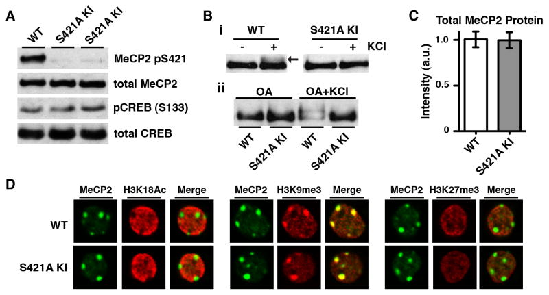
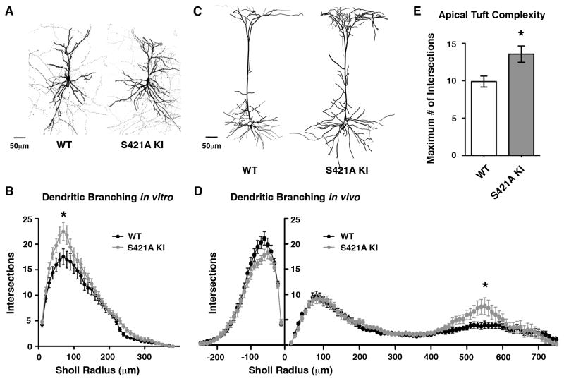
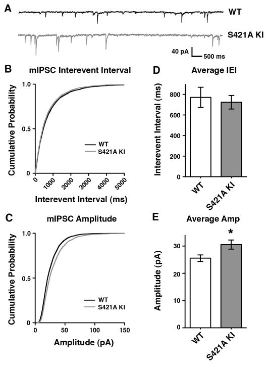
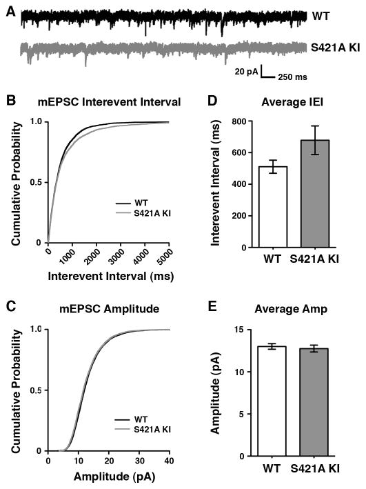
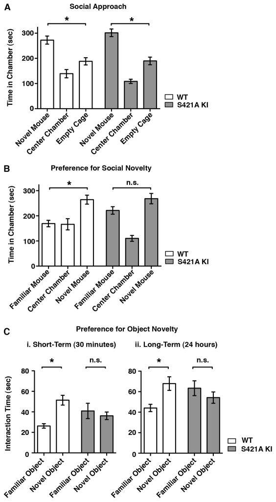
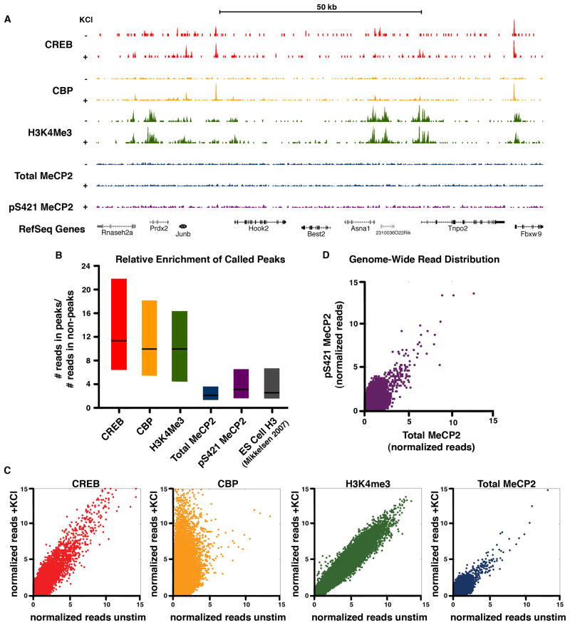
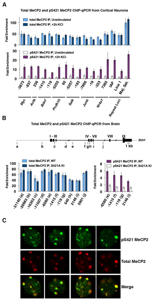
Autism spectrum disorders such as Rett syndrome (RTT) have been hypothesized to arise from defects in experience-dependent synapse maturation. RTT is caused by mutations in MECP2, a nuclear protein that becomes phosphorylated at S421 in response to neuronal activation. We show here that disruption of MeCP2 S421 phosphorylation in vivo results in defects in synapse development and behavior, implicating activity-dependent regulation of MeCP2 in brain development and RTT. We investigated the mechanism by which S421 phosphorylation regulates MeCP2 function and show by chromatin immunoprecipitation-sequencing that this modification occurs on MeCP2 bound across the genome. The phosphorylation of MeCP2 S421 appears not to regulate the expression of specific genes; rather, MeCP2 functions as a histone-like factor whose phosphorylation may facilitate a genome-wide response of chromatin to neuronal activity during nervous system development. We propose that RTT results in part from a loss of this experience-dependent chromatin remodeling.


Figures

Figure 1
Figure 1. Selective Loss of the Activity-Dependent Phosphorylation of MeCP2 S421 in vivo Preserves the Expression Level and Localization of MeCP2
(A) Western blot analysis of nuclear extracts prepared from the forebrains of two 4-week-old male MeCP2 S421A knock-in mice (S421A KI) and their wild-type (WT) littermate using antibodies specific to MeCP2 pS421, and total MeCP2. Parrallel blots for pS133 CREB and total CREB show that there is no gross disruption of activity-dependent signaling in the brain. (B) Anti-total MeCP2 Western blots of lysates from E16 + 5 DIV dissociated hippocampal neurons derived from MeCP2 S421A knock-in mice or wild-type littermates. (i) Arrow indicates the S421 phosphorylation-dependent slowly-migrating form of MeCP2 induced by 60 minute 55 mM KCl stimulation in wild-type cells (ii) 30 minute pre-treatment with okadaic acid (0.25 μM) to inhibit protein phosphatase activity increases the proportion of slowly-migrating MeCP2 induced by the 60 minute membrane depolarization. MeCP2 S421A neurons do not display this slowly-migrating form of MeCP2 in response to membrane depolarization. (C) Quantification of Western blot analysis for the forebrains of 4-week-old MeCP2 S421A mice (n=3) and their wild-type littermates (n=3) using anti-total MeCP2 antiserum. Data are mean ± SEM. See also Figure S1. (D) Immunocytochemistry of E16 + 22 DIV cortical neurons derived from MeCP2 S421A mice and wild-type littermates using an antibody against total MeCP2 and costaining with anti-histone H3 lysine 18 acetylation (H3K18Ac, active chromatin), anti-histone H3 lysine 9 trimethylation (H3K9me3, heterochromatin) or anti-histone H3 lysine 27 trimethylation (H3K27me3, repressed chromatin). Single 63X confocal planes of representative neuronal nuclei are shown.
Figure 2
Figure 2. Increased Dendritic Complexity Upon Loss of MeCP2 S421 Phosphorylation
(A) Dissociated E16+21–22 DIV cortical neurons from MeCP2 S421A knock-in mice or wild-type littermates transfected with GFP to visualize dendritic morphology. Maximal intensity projections of a series of confocal planes for representative 25X images are shown. (B) Sholl analysis measuring the number of dendritic branch points at 10 μm radial increments from the cell soma reveal an increase in dendritic branching in the MeCP2 S421A cells (p < 0.05, two-way repeated measures ANOVA) due to increased branch points 60–80 μm from the cell soma (p < 0.01, Bonferroni posttests). Wild-type (n=26) and MeCP2 S421A knock-in (n=33) neurons from two independent experiments. (C) Representative images of single layer V pyramidal neurons expressing GFP under from the Thy1 promoter in 4-week-old forebrains of GFP-M transgenic mice. Tracings are shown of maximal intensity projections of a series of 10X confocal planes taken from coronal cortical sections. (D) Sholl analysis of dendritic branch points at 10 μm radial increments from the cell soma reveals an increase in dendritic branching of MeCP2 S421A layer V pyramidal neuron apical dendrites 530 (p < 0.05), 540 (p < 0.01), 550 (p < 0.01) and 560 μm (p < 0.05) from the cell soma (two-way repeated measures ANOVA with Bonferroni posttests). Wild-type (n=29) and MeCP2 S421A knock-in (n=16) neurons from three independent littermate pairs. (E) The maximum number of dendritic intersections ≥ 400 μm from the cell soma observed by Sholl analysis of each GFP-M+ layer V pyramidal cell in MeCP2 WT and S421A knock-in mice. The increase in maximum number of intersections in the MeCP2 S421A knock-in cells compared to WT indicates that the MeCP2 S421 knock-in apical tuft is more complex regardless of the exact distance of the apical tuft from the cell soma. p < 0.01 by Student’s t-test. All data shown in Figure 2 are mean ± SEM.
Figure 3
Figure 3. Loss of Activity-Dependent MeCP2 S421 Phosphorylation Regulates Development of Inhibitory Synaptic Transmission in vivo
(A) Representative traces of mIPSCs recorded from layer II/II V1 pyramidal neurons in acute cortical slices from P16–17 MeCP2 S421A knock-in mice or their wild-type littermates. (B–C) Cumulative probability distribution of mIPSC interevent intervals (B) and amplitudes (C) recorded from MeCP2 S421A or wild-type littermates. (D–E) Average interevent interval (D) and amplitude (E) of mIPSCs recorded from wild-type or MeCP2 S421A neurons. Data are mean ± SEM. The difference in amplitude is statistically significant, p < 0.05 by Student’s t-test. Data shown represent 250 random events drawn from each of the wild-type (n=19) or MeCP2 S421A knock-in (n=21) cells analyzed, recorded from mice from 6 independent litters.
Figure 4
Figure 4. Analysis of mEPSCs in the MeCP2 S421A Knock-In Mouse
(A) Representative traces of mEPSCs recorded from layer II/II V1 pyramidal neurons in P16–17 MeCP2 S421A knock-in mice or their wild-type littermates. (B–C) Cumulative probability distributions of mEPSC interevent intervals (B) or amplitudes (C) recorded from MeCP2 S421A or wild-type littermates. (D–E) Average interevent interval (D) and ampltitude (E) of mEPSCs recorded from MeCP2 S421A or wild-type neurons. Data are mean ± SEM, p = 0.1 by Student’s t-test. Data shown represent 250 random events drawn from each of the wild-type (n=23) or MeCP2 S421A knock-in (n=25) cells analyzed, recorded from 6 pairs of littermates.
Figure 5
Figure 5. Defects in Behavioral Response to Novel Experience in MeCP2 S421A Knock-In Mice
(A) Sociability is not affected by loss of MeCP2 S421 phosphorylation. Behavior of wild-type (n = 24) and MeCP2 S421A knock-in (n=21) mice in a three-chamber apparatus with a single novel mouse placed in a small wire cage on one of the side chambers. Data shown are time spent in each chamber over the 10 minute trial period. Wild-type and S421A mice spent significantly more time in the chamber with the novel mouse (WT p < 0.001, S421A p < 0.01). In this assay wild-type and MeCP2 S421A mice were statistically indistinguishable. (B) MeCP2 S421A mice do not lose interest in familiar mice. Behavior of wild-type (n = 24) and MeCP2 S421A knock-in (n=21) mice in a three-chamber apparatus with a familiar mouse placed in one of the side chambers and a novel mouse placed in the opposite side chamber. Time spent in each chamber over the 10 minute trial period is shown. Wild-type mice spent significantly more time with the novel mouse than with the familiar mouse or alone (p < 0.01). The amount of time spend with each test mouse did not differ significantly for MeCP2 S421A knock-in mice. (C) MeCP2 S421A mice do not distinguish between familiar and novel objects. Behavior of wild-type (n = 9) and MeCP2 S421A (n = 9) mice when placed in an arena with a familiar inanimate object and a novel inanimate object placed at opposite ends. In the short-term assay (i), the familiar object was first introduced 30 minutes prior to the trial. In the long-term assay (ii), the familiar object was first introduced 24 hours prior to the trial. Wild-type mice spend significantly more time interacting with the novel object than with the familiar object in both assays (i, p < 0.01; ii, p < 0.05), while MeCP2 S421A knock-in mice do not. Total time spent exploring each object over a 10 minute trial period is shown for each assay. Data for all plots are mean ± SEM, p-values from one-way ANOVA with Bonferroni multiple comparison correction.
Figure 6
Figure 6. ChIP-Seq analysis reveals a histone-like binding profile for total MeCP2 and global phosphorylation of MeCP at S421 upon neuronal stimulation
(A) ChIP-Seq profiles for total and pS421 MeCP2 as well as CREB, CBP and H3K4me3 (Kim et al., 2010) across a representative genomic region. ChIP samples were isolated from E16 + 7 DIV cortical cultures, unstimulated (‘−‘) or stimulated for 2 hr with 55mM KCl (‘+’). The location of RefSeq genes, including the activity-regulated immediate-early gene Junb, are indicated. (B) Box plots (1st quartile, median, and 3rd quartile) displaying the ratio of the number of reads found at called peaks versus non-peak control regions are shown for each ChIP-seq data set (2hr KCl stimulated). Enrichment for pan-histone H3 ChIP-Seq peaks from embryonic stem cells is shown for comparison (H3ES, Mikkelsen et al., 2008). (C) Scatter plots depict normalized ChIP-Seq reads from E16 + 7 DIV cortical cultures for 1000 bp windows tiled across the genome before and after stimulation (2hr KCl). Data points on the diagonal indicate no substantial change in ChIP signal upon stimulation. Points located off the diagonal, as seen for CBP, indicates dynamic change in response to neuronal activity. (D) Scatter plot depicts normalized ChIP-Seq reads of pS421 MeCP2 versus total MeCP2 from stimulated dissociated cortical cultures (E16 + 7 DIV, 2 hr KCl) for 1000 bp windows tiled across the genome. Distribution along the diagonal indicates that the profile of pS421 reads tracks with total MeCP2 reads across the genome.
Figure 7
Figure 7. ChIP-qPCR and immunohistochemistry confirm broad binding of total MeCP2 and global MeCP2 S421 phosphorylation in vitro and in vivo
(A) ChIP-qPCR analysis of total MeCP2 (top panel) and pS421 MeCP2 (bottom panel) in cultured cortical neurons (E16 + 7 DIV) that were either membrane depolarized for 2 hours (55mM KCl) or left unstimulated. (B) Total MeCP2 (left) and pS421 MeCP2 (right) ChIP-qPCR from the forebrains of 7-week-old MeCP2 S421A mice or their wild-type littermates. The location of PCR amplicons across the Bdnf locus are shown in diagram as a-j. Nucleotide position is given relative to start of the activity-dependent Bdnf promoter IV. All ChIP data is presented as fold enrichment above a negative control ChIP performed in parallel on each sample using the same antiserum that had been preincubated with the peptide antigen to which it was raised. Data are mean ± SEM from three independent experiments. (C) Immunocytochemistry of nuclei from wild-type E16 + 10 DIV mouse cortical neurons following 30 min membrane depolarization with 55 mM KCl. Antibodies used are specific for total MeCP2 or pS421 MeCP2 (see also Figure S4). Representative 63X images from single confocal planes are shown.

Comment in

References

    1. Armstrong DD. Neuropathology of Rett syndrome. Mental Retardation and Developmental Disabilities Research Reviews. 2002;8:72–76. - PubMed
    1. Ballas N, Lioy DT, Grunseich C, Mandel G. Non cell autonomous influence of MeCP2-deficient glia on neuronal dendritic morphology. Nature Neuroscience. 2009;12:311–317. - PMC - PubMed
    1. Chahrour M, Jung SY, Shaw C, Zhou X, Wong STC, Qin J, Zoghbi HY. MeCP2, a Key Contributor to Neurological Disease, Activates and Represses Transcription. Science. 2008;320:1224–1229. - PMC - PubMed
    1. Chahrour M, Zoghbi H. The Story of Rett Syndrome: From Clinic to Neurobiology. Neuron. 2007;56:422–437. - PubMed
    1. Chao HT, Zoghbi HY, Rosenmund C. MeCP2 Controls Excitatory Synaptic Strength by Regulating Glutamatergic Synapse Number. Neuron. 2007;56:58–65. - PMC - PubMed

Publication types

MeSH terms