. 2011 Oct 6;72(1):101-10.
doi: 10.1016/j.neuron.2011.07.030.

Rod vision is controlled by dopamine-dependent sensitization of rod bipolar cells by GABA

Affiliations

Rod vision is controlled by dopamine-dependent sensitization of rod bipolar cells by GABA

Rolf Herrmann et al. Neuron. .

Abstract

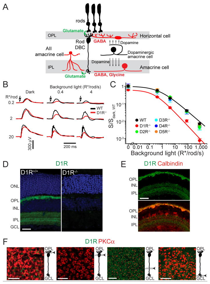
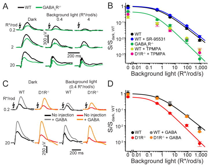
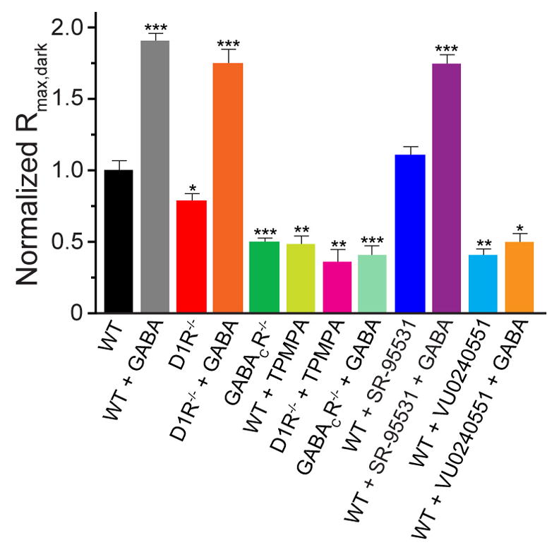
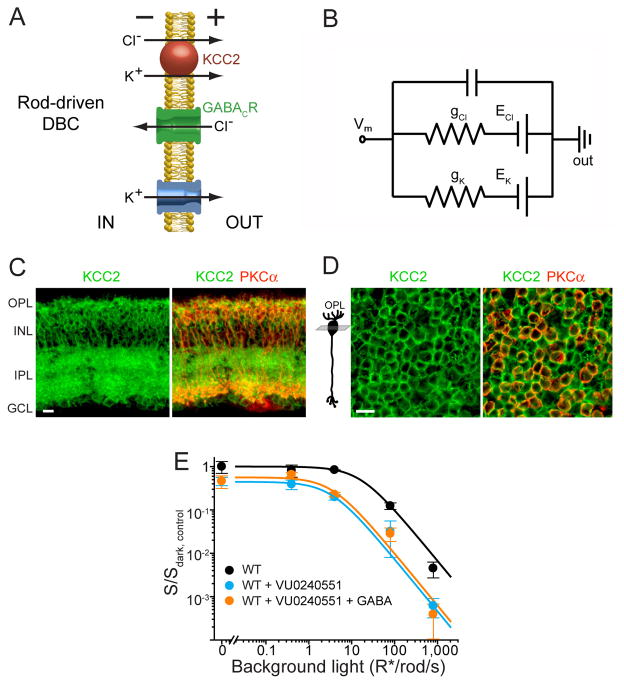
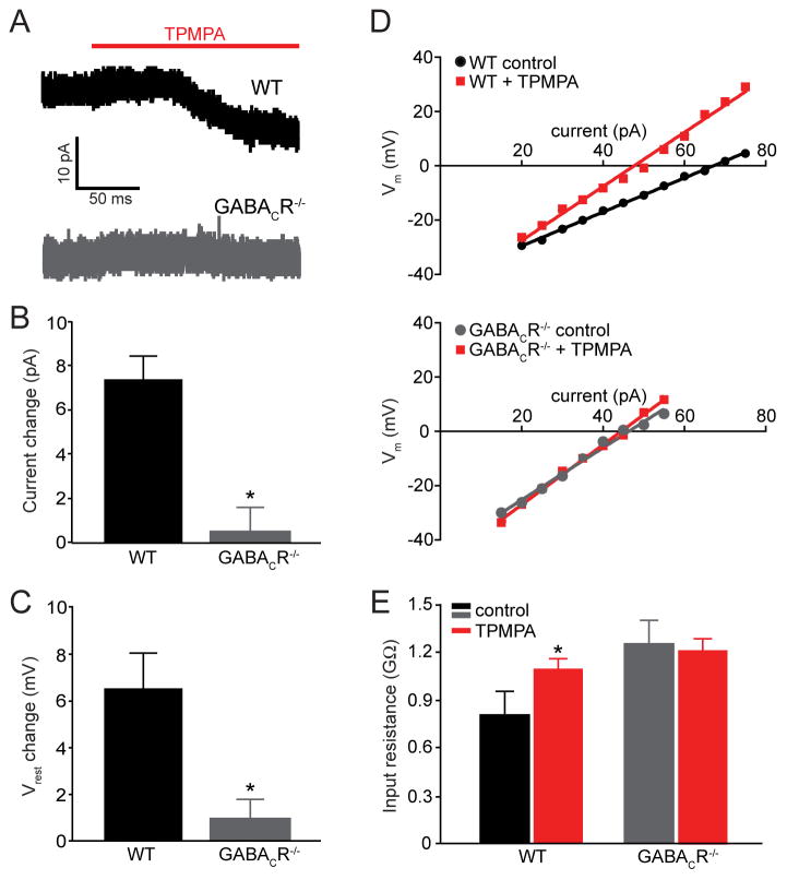
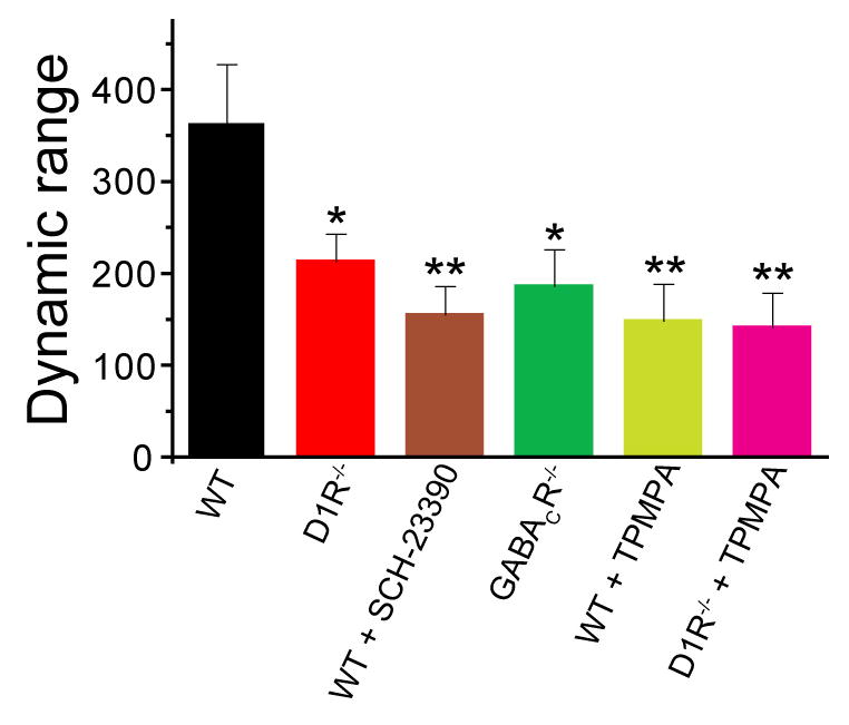
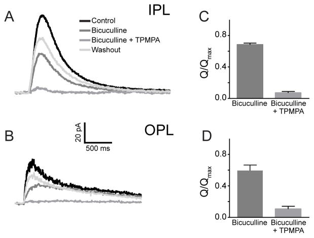
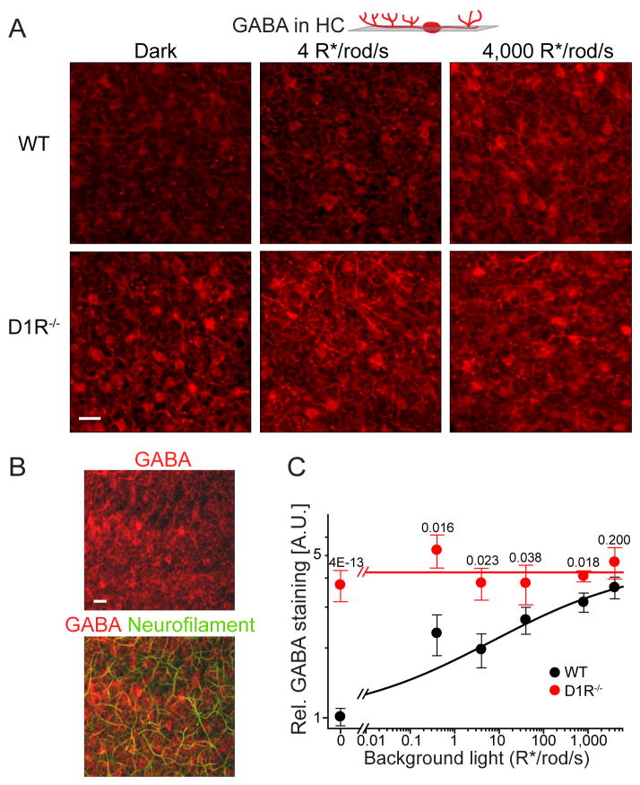
Dark and light adaptation of retinal neurons allow our vision to operate over an enormous light intensity range. Here we report a mechanism that controls the light sensitivity and operational range of rod-driven bipolar cells that mediate dim-light vision. Our data indicate that the light responses of these cells are enhanced by sustained chloride currents via GABA(C) receptor channels. This sensitizing GABAergic input is controlled by dopamine D1 receptors, with horizontal cells serving as a plausible source of GABA release. Our findings expand the role of dopamine in vision from its well-established function of suppressing rod-driven signals in bright light to enhancing the same signals under dim illumination. They further reveal a role for GABA in sensitizing the circuitry for dim-light vision, thereby complementing GABA's traditional role in providing dynamic feedforward and feedback inhibition in the retina.


Figures

Figure 1
Figure 1. Reduced sensitivity and operational range of rod-driven DBCs in D1R−/− mice and localization of D1R in the retina
(A) Cartoon illustrating the cell types discussed in this study. See text for details. (B) Representative ERG recordings from WT and D1R−/− mice under dark- and light-adapted conditions. Light intensity for flash and background light is given in units of photoexcited rhodopsin molecules per rod (R*/rod) and photoexcited rhodopsin molecules per rod per second (R*/rod/s), respectively. (C) Sensitivities of rod-driven ERG b-waves were determined for five dopamine receptor knockout mice, normalized to the dark sensitivity of WT mice (S/Sdark, WT) and plotted as a function of background light intensity (mean ± SEM). Here and in figures below see Table S1 for the summary of fitting parameters. (D) D1R immunostaining in WT and D1R−/− retinal cross-sections (green). Nuclei are stained with Hoechst (blue). (E) WT retinal cross-section stained for D1R and calbindin, which labels horizontal cells in the OPL/INL and a class of amacrine cells in the INL/IPL. (F) Confocal tangential z-sections of a retina flat-mount co-stained for D1R and the rod DBC marker, PKCα. Positions of individual z-sections relative to DBC morphology are illustrated in cartoons on the right from each panel. Abbreviations: ONL – outer nuclear layer; OPL – outer plexiform layer; INL – inner nuclear layer; IPL – inner plexiform layer; GCL – ganglion cell layer. Scale bars: 25 μm.
Figure 2
Figure 2. The sensitivity and operational range of rod-driven DBC responses are regulated by GABACR
(A) ERG recordings from WT and GABACR−/− mice under dark- and light-adapted conditions. (B) ERG b-wave sensitivity plots for the following mice and conditions: WT, GABACR−/−, WT injected with the GABAAR antagonist SR-95531, WT injected with GABACR antagonist TPMPA, D1R−/− injected with TPMPA. (C) ERG recordings from dark- or light-adapted WT and D1R−/− mice with and without GABA injections. (D) WT and D1R−/− ERG b-wave sensitivity plots in the absence or presence of injected GABA.
Figure 3
Figure 3. Normalized maximal dark-adapted amplitudes of rod-driven b-waves differ across mice with and without GABACR-meditated input
Paired t-tests between WT and each other animal/condition yielded p-values <0.025 (*), <0.005 (**) and <0.001 (***).
Figure 4
Figure 4. Rod-driven DBC responses are sensitized by GABACR-dependent chloride influx
(A) Cartoon illustrating the role of KCC2 in chloride extrusion from rod DBCs and the roles of chloride and potassium fluxes in tonic rod DBC hyperpolarization. (B) Equivalent circuit illustrating exchangeability and additivity of chloride and potassium conductances in creating the driving force for light-induced cation influx resulting in DBC depolarization. Under these conditions, the resting potential is defined as Vm = (gK·EK + gCl·ECl)/(gK + gCl), where gK and gCl are potassium and chloride conductances, and EK and ECl are potassium and chloride reversal potentials, respectively. (C) Retinal cross-section from a WT mouse immunostained for KCC2 and the rod DBC marker, PKCα (see Figure 1 for abbreviations of retina layers). (D) Confocal tangential z-section through bipolar cell somata of a flat-mounted retina co-stained for KCC2 and PKCα. Position of the z-section relative to DBC morphology is shown in the cartoon on the left. Scale bars in (C) and (D): 10 μm. (E) ERG b-wave sensitivity plots for WT mice with and without injections of the KCC2 antagonist VU0240551, or with co-injection of VU0240551 and GABA. Ethanol (95%), the vehicle for VU0240551, was injected in WT eyes as a control; the presence of ethanol shifted the sensitivity plot for WT mice, but the relative reduction in sensitivity in GABACR−/− vs. WT and in KCC2 blockade vs. WT+ethanol was similar.
Figure 5
Figure 5. WT rod DBCs have a GABACR-mediated tonic current that hyperpolarizes their resting potential and decreases their input resistance
(A) Representative currents in WT and GABACR−/− rod DBCs in the presence of 5 μM GABA evoked by bath application of 50 μM TPMPA. (B) The average change in tonic current caused by TPMPA in WT (n=7) and GABACR−/− rod DBCs (n=6) differed significantly (p=0.001). (C) −/− rod DBCs (n=5; TPMPA depolarizes the resting potential (Vrest) of WT (n=8) but not GABACR p =0.03). (D) The linear range of current-voltage relationships of representative WT and GABACR−/− rod DBCs in the presence and absence of 50 μM TPMPA. A change in slope was observed in 6 out of 8 WT cells and in none of the 5 GABACR−/− rod DBCs. (E) Input resistances of WT rod DBCs were increased in the presence of TPMPA, but were not changed in GABACR−/− rod DBCs. The input resistances calculated from the slopes of current-voltage curves were: 0.8 ± 0.2 GΩ and 1.1 ± 0.08 GΩ (mean ± SEM, p = 0.03) for WT rod DBCs in the absence and presence of TPMPA, respectively; 1.26 GΩ ± 0.4 and 1.20 ± 0.07 GΩ for GABACR−/− rod DBCs in the absence and presence of TPMPA, respectively. The input resistance of WT rod DBCs in the presence of TPMPA was similar to GABACR−/− rod DBCs in the presence or absence of TPMPA.
Figure 6
Figure 6. Knockout or blockade of D1R and/or GABACR reduces the dynamic range of ERG b-wave responses
The response dynamic range for each animal/condition (i.e. the range of intensities covering between 5% to 95% of the maximal b-wave response) was calculated as 102.56/n (Thibos and Werblin, 1978), where n is the Hill coefficient for the rod-driven b-wave component calculated from fitting b-wave stimulus-response curves to Equation 1 (see Supplementary Experimental Procedures). The data were averaged from all recording sets obtained in the dark and in the presence of background light. Paired t-tests between WT and each other animal/condition yielded p-values of <0.05 (*) and <0.01 (**).
Figure 7
Figure 7. GABAergic currents are mediated by GABAA and GABAC receptors in both dendrites and axon terminals of rod DBCs
(A) and (B) Representative current responses to brief puffs of GABA delivered focally onto the axon terminals in the IPL (A) or dendrites in the OPL (B) of the same rod DBC in the presence or absence of the GABAAR antagonist bicuculline (500 μM) or a mixture of bicuculline with the GABACR antagonist TPMPA (50 μM). (C) and (D) Averaged normalized charge transfer (Q/Qmax ) of rod DBCs in response to GABA puffs in the IPL and OPL and in the presence of bicuculline alone or bicuculline +TPMPA. In both cases, bicuculline reduced responses to 80% of the control in the IPL and 66% in the OPL (n=6). TPMPA + bicuculline essentially eliminated responses in both IPL and OPL (7% vs 12% in IPL vs. OPL; n=6). An ANOVA compared the reduction in these responses from control. Both OPL and IPL responses were significantly different in the presence of bicuculline as well as bicuculline + TPMPA (p < 0.001). There was no difference when similar conditions were compared between IPL and OPL.
Figure 8
Figure 8. Horizontal cells serve as a putative site of D1R-regulated GABA release
(A) GABA immunostaining of horizontal cells in single tangential confocal sections of WT and D1R−/− retinas. The position of optical sections is illustrated on the cartoon above. Animals were light-conditioned as indicated in the panel. (B) GABA co-immunostaining with neurofilaments in a single tangential confocal section through the horizontal cell layer in WT retina. (C) Quantification of GABA immunostaining in horizontal cells from mice subjected to different levels of background illumination (mean ± SEM; 3 to 6 retinas were analyzed for each condition; p-values for the difference between animal types at the same condition are shown on the top of each pair).

Similar articles

Cited by

References

    1. Amin J, Weiss DS. Homomeric rho 1 GABA channels: activation properties and domains. Receptors Channels. 1994;2:227–236. - PubMed
    1. Babai N, Thoreson WB. Horizontal cell feedback regulates calcium currents and intracellular calcium levels in rod photoreceptors of salamander and mouse retina. J Physiol. 2009;587:2353–2364. - PMC - PubMed
    1. Carlsson A, Waters N, Holm-Waters S, Tedroff J, Nilsson M, Carlsson ML. Interactions between monoamines, glutamate, and GABA in schizophrenia: new evidence. Annu Rev Pharmacol Toxicol. 2001;41:237–260. - PubMed
    1. Chavez AE, Grimes WN, Diamond JS. Mechanisms underlying lateral GABAergic feedback onto rod bipolar cells in rat retina. J Neurosci. 2010;30:2330–2339. - PMC - PubMed
    1. Deniz S, Wersinger E, Schwab Y, Mura C, Erdelyi F, Szabo G, Rendon A, Sahel JA, Picaud S, Roux MJ. Mammalian retinal horizontal cells are unconventional GABAergic neurons. J Neurochem. 2011;116:350–362. - PubMed

Publication types

MeSH terms