. 2011 Oct 25;21(20):1685-94.
doi: 10.1016/j.cub.2011.08.049. Epub 2011 Oct 6.

The tubulin deglutamylase CCPP-1 regulates the function and stability of sensory cilia in C. elegans

Affiliations

The tubulin deglutamylase CCPP-1 regulates the function and stability of sensory cilia in C. elegans

Robert O'Hagan et al. Curr Biol. .

Abstract

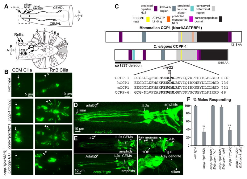
Background: Posttranslational modifications (PTMs) such as acetylation, detyrosination, and polyglutamylation have long been considered markers of stable microtubules and have recently been proposed to guide molecular motors to specific subcellular destinations. Microtubules can be deglutamylated by the cytosolic carboxypeptidase CCP1. Loss of CCP1 in mice causes cerebellar Purkinje cell degeneration. Cilia, which are conserved organelles that play important diverse roles in animal development and sensation, contain axonemes comprising microtubules that are especially prone to PTMs.

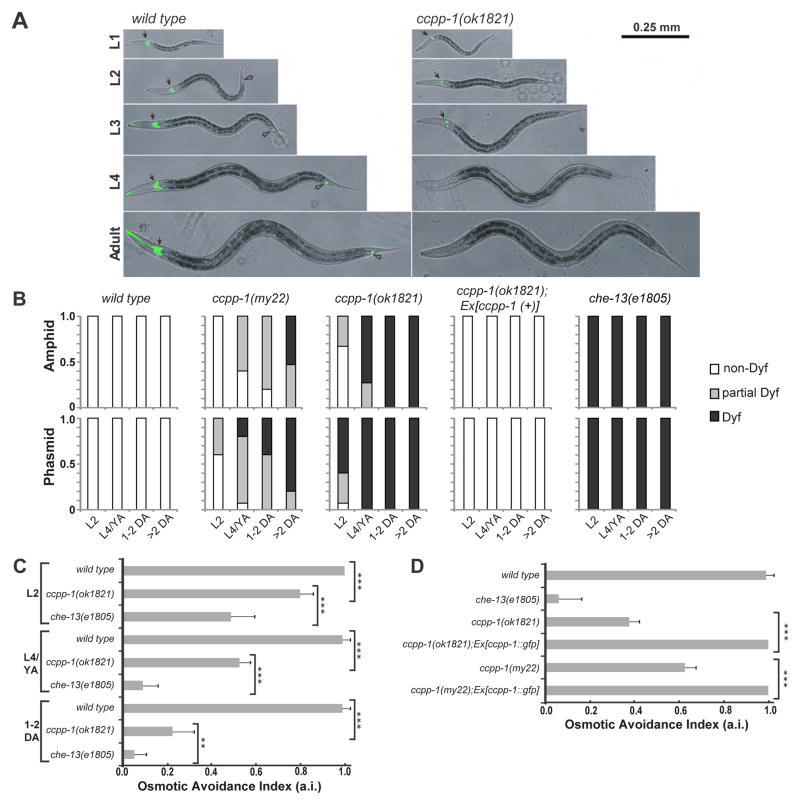
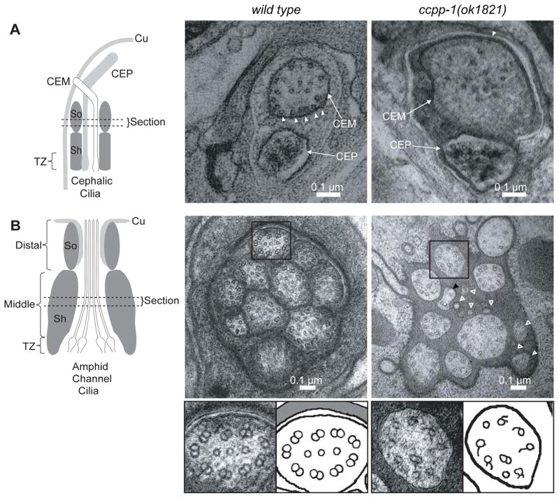
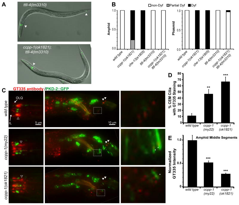
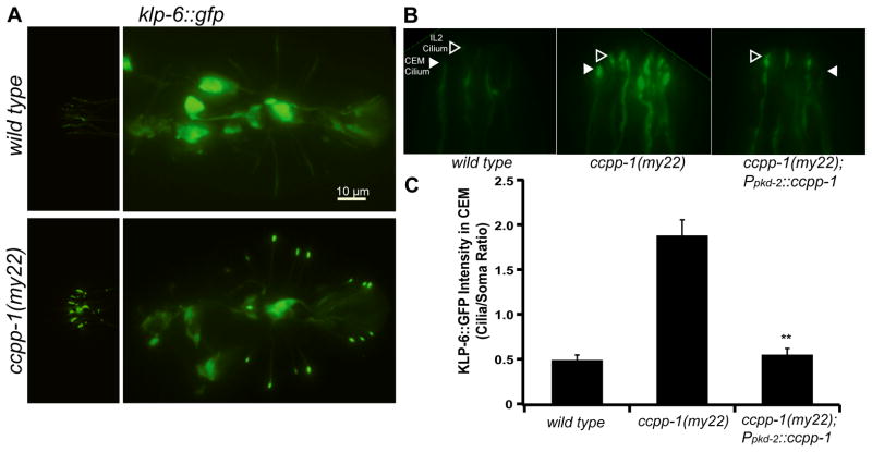
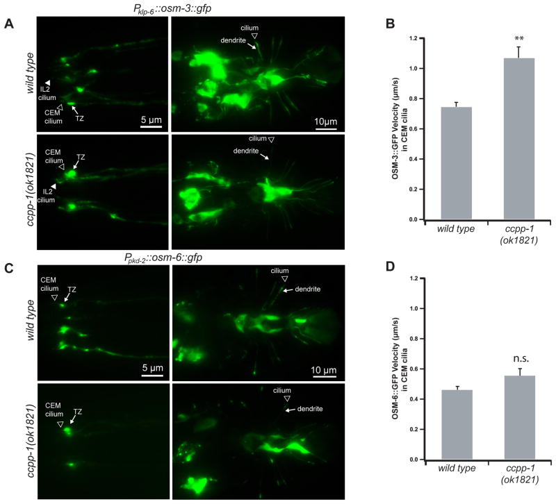
Results: Here, we report that a CCP1 homolog, CCPP-1, regulates the ciliary localization of the kinesin-3 KLP-6 and the polycystin PKD-2 in male-specific sensory neurons in C. elegans. In male-specific CEM (cephalic sensilla, male) cilia, ccpp-1 also controls the velocity of the kinesin-2 OSM-3/KIF17 without affecting the transport of kinesin-II cargo. In the core ciliated nervous system of both males and hermaphrodites, loss of ccpp-1 causes progressive defects in amphid and phasmid sensory cilia, suggesting that CCPP-1 activity is required for ciliary maintenance but not ciliogenesis. Affected cilia exhibit defective B-tubules. Loss of TTLL-4, a polyglutamylating enzyme of the tubulin tyrosine ligase-like family, suppresses progressive ciliary defects in ccpp-1 mutants.

Conclusions: Our studies suggest that CCPP-1 acts as a tubulin deglutamylase that regulates the localization and velocity of kinesin motors and the structural integrity of microtubules in sensory cilia of a multicellular, living animal. We propose that the neuronal degeneration caused by loss of CCP1 in mammals may represent a novel ciliopathy in which cilia are formed but not maintained, depriving the cell of cilia-based signal transduction.


Figures

Fig. 1
Fig. 1. CCPP-1 is required for PKD-2 localization and is expressed in the ciliated sensory nervous system
A Diagram of the male-specific CEM neurons in the head, and HOB and RnB neurons in the tail. Box illustrates the region of CEM cilia and distal dendrites shown in the epifluorescent images. In the ventral-up cartoon of the male tail, the RnBs innervate the tail rays; R3B dendrite is shown as an example. B In wild-type males, PKD-2::GFP faintly illuminated cilia of the CEM neurons, the HOB cilium, and the cilia of the ray B-type neurons (RnB, where n = 1 – 9, excluding 6). ccpp-1(my22) and ccpp-1(ok1821) mutants exhibit the Cil phenotype. Arrows point to CEM cilia; arrowheads point to R1B and R2B cilia. Expression of genomic ccpp-1 rescued the Cil phenotype of ccpp-1(ok1821). C CCPP-1 contains several conserved regions, including a zinc carboxypeptidase domain, an aspartic-acid rich domain, a conserved “NT” region [12], and predicted NLS (nuclear localization signal; [35]). CCPP-1 also contains a predicted leucine zipper near the N-terminus (ScanProsite, [36]). The my22 lesion affects the conserved sequence FESGNL in the NT region of CCPP-1. D In adult hermaphrodites, CCPP-1::GFP expression was expressed in amphid and IL2 core ciliated sensory neurons. Cilium containing CCPP-1::GFP is indicated. E Confocal projections of CCPP-1::GFP expression in males. Top (L4): CCPP-1::GFP was expressed in (left) amphid, IL2, and CEM neurons and (right) male tail ray and HOB neuronal cell bodies (empty arrowheads). Gubernacular erector (g.e.) and retractor (g.r.) muscles are indicated. Bottom (1 day old adult): CCPP-1::GFP localization in ray neuron dendrites (arrow) and a cilium (arrowhead). F ccpp-1 mutants are defective in response behavior. Number of trials and number of males for each genotype was as follows: wild type, 7 trials, N = 70 males; ok1821, 6 trials, N = 60; ok1821;Ex[ccpp-1(+)], 3 trials, N = 29; ok1821;Ex[ccpp-1::gfp], 3 trials, N = 24; my22, 4 trials, N = 40; my22;Ex[ccpp-1::gfp], 5 trials, N = 39. Error bars indicate SEM; **indicates ccpp-1 mutants were statistically different (p < 10−4, ANOVA/Tukey HSD test) from mutants expressing ccpp-1 transgenes, which were similar to wild type. See also Fig. S1.
Fig. 2
Fig. 2. ccpp-1 mutants exhibited progressive Dyf and Osm defects
A Amphid (solid arrows) and phasmid (hollow arrows) ciliated neurons of wild type hermaphrodites were stained by DiI (pseudocolored green) at all developmental stages. ccpp-1(ok1821) amphid cilia in L1 larvae stained normally, but became Dyf in later larval stages and adults. B Dyf defects were scored in amphid and phasmid neurons in wild-type, my22, ok1821, and che-13(e1805) young adults (24 hours post-L4). 15 animals were scored for each stage/genotype. C Wild-type animals exhibit osmotic avoidance behavior when challenged with an 8 M glycerol ring. 80 hermaphrodites (8 trials, 10 per trial) were tested for each stage/genotype. Osmotic avoidance index (a.i.) is the fraction of animals that avoided crossing the ring. D The ccpp-1 Osm defect was rescued by the ccpp-1::gfp transgene (8 trials, 10 animals per trial tested). (Error bars indicate SEM; ** indicates p = 0.0022 with Fischer’s Exact Test; *** indicates p < 0.0001 with Fischer’s Exact Test).
Fig. 3
Fig. 3. ccpp-1(ok1821) mutants exhibit ciliary ultrastructure defects
Left diagrams show regions from which the cephalic (CEM and CEP) and amphid cilia images were taken (Cu = cuticle; Sh = sheath cell; So = socket cell; TZ =transition zone). A EM Images of CEM and CEP cilia in wild-type and ccpp-1(ok1821) mutant adult males. Wild-type CEM cilia image (taken from a tomogram) contained many singlet MTs (19 singlets in section shown) closely apposed to the membrane (arrowheads). ok1821 CEM cilia had fewer singlets (15 singlets in section shown), which were more distant from the membrane (arrowhead indicates one singlet near membrane). The ok1821 CEM cilium diameter was larger than wild type. B Thin sections of amphid cilia in wild-type and ccpp-1(ok1821) adult males. Wild-type middle segments contain ten axonemes, each of which typically has nine outer doublets plus a variable number of inner singlets. The ok1821 middle segment contained only eight intact axonemes plus what appear to be fragments of two cilia (hollow white arrowheads), one of which contains a singlet with attached broken B-tubule (black arrowhead). Most ok1821 mutant axonemes had fewer MTs, with many doublets replaced by singlets or broken B-tubules. Bottom, boxed wild-type and mutant axonemes accompanied by cartoons. Refer to Table 1 for quantification of images.
Fig. 4
Fig. 4. Loss of CCPP-1 results in altered polyglutamylation of sensory cilia
A Dye uptake (pseudocolored green) was normal in young adult hermaphrodite ttll-4(tm3310) mutants (left), which were previously reported to lack polyglutamylation in cilia [21]. Deletion of ttll-4 suppressed the Dyf phenotype of ccpp-1 (right). Arrowheads indicate dye-filled amphid and phasmid neurons. B Penetrance of Dyf defects in amphid and phasmid neurons. 50 young adult hermaphrodites per genotype were tested. C Staining of wild-type and ccpp-1 mutant young adult males with GT335, a monoclonal antibody that detects polyglutamylation, most prominently in amphid middle segments (hollow arrowhead). An IL and putative OLQ cilium are indicated in the nose. GT335 rarely stained wild-type CEM cilia, which express PKD-2::GFP. In the tail, phasmid cilia (solid arrowheads) were brightly stained. Right, enlarged boxed area containing several ray neuron dendrites and cilia. Asterisks mark polyglutamylation signals that are abnormally localized to the ciliary base in ccpp-1 mutants. D ccpp-1 mutations increased the incidence of GT335 staining of CEM cilia, which were identified by PKD-2::GFP (** indicates P < 0.01, *** indicates P < 0.001 vs. wild type, ANOVA/Tukey test; N = 19 – 22 males per genotype; scored blindly). E The normalized peak pixel value of GT335 staining in the area containing amphid middle segments was significantly lower in mutants (Error bars indicate SEM; *** indicates p < 10−5 versus wild type, by ANOVA/Tukey HSD test; N = 10 males per genotype). See also Fig. S2.
Fig. 5
Fig. 5. CCPP-1 is needed for proper localization of KLP-6::GFP in IL2 and male-specific neurons
A KLP-6::GFP localization in IL2 and CEM neurons (left) and in HOB and RnB neurons in the tail (right) is diffuse in wild-type males. In contrast, KLP-6::GFP is highly enriched in cilia in ccpp-1(my22) mutants. B A magnified view of IL2 (hollow arrowheads) and CEM (solid arrowheads) cilia containing KLP-6::GFP in wild-type, ccpp-1(my22), and ccpp-1(my22) rescued young adult males. C Quantification of KLP-6::GFP fluorescence in cilia/somata of CEM neurons. KLP-6::GFP localization defects in ccpp-1(my22) mutant cilia were rescued by Ppkd-2::ccpp-1 in male specific CEM (and RnB, not shown) neurons (Error bars indicate SEM; N = 9 – 10 animals per genotype; ** indicates P < 0.01 by t-test versus ccpp-1(my22).)
Fig. 6
Fig. 6. CCPP-1 regulates the velocity of OSM-3::GFP but not kinesin-II-driven IFT-B polypeptide OSM-6::GFP in CEM cilia
A In wild-type and ccpp-1(ok1821) young adult males, klp-6 promoter-driven OSM-3::GFP was visible diffusely in cell bodies, dendrites, and cilia, with some accumulation in transition zones (TZ). B OSM-3::GFP particles moved faster in ccpp-1 CEM cilia (78 particles in 7 wild-type males; 81 particles in 10 ccpp-1(ok1821) males; ** indicates p < 10−4 by ANOVA/Tukey test). C, D OSM-6::GFP localization and velocity in CEM cilia was similar in wild-type and ok1821 males (Error bars indicate SEM; 55 particles in 6 wild-type males; 61 particles in 7 ccpp-1(ok1821) males; n.s. indicates no significant difference). See also Fig. S3.

References

    1. Fliegauf M, Benzing T, Omran H. When cilia go bad: cilia defects and ciliopathies. Nat Rev Mol Cell Biol. 2007;8:880–893. - PubMed
    1. Scholey JM. Intraflagellar transport motors in cilia: moving along the cell’s antenna. J Cell Biol. 2008;180:23–29. - PMC - PubMed
    1. Pan X, Ou G, Civelekoglu-Scholey G, Blacque OE, Endres NF, Tao L, Mogilner A, Leroux MR, Vale RD, Scholey JM. Mechanism of transport of IFT particles in C. elegans cilia by the concerted action of kinesin-II and OSM-3 motors. J Cell Biol. 2006;174:1035–1045. - PMC - PubMed
    1. Morsci NS, Barr MM. Kinesin-3 KLP-6 Regulates Intraflagellar Transport in Male-Specific Cilia of Caenorhabditis elegans. Curr Biol. 2011;21:1239–1244. - PMC - PubMed
    1. Bulinski JC, Gundersen GG. Stabilization of post-translational modification of microtubules during cellular morphogenesis. BioEssays : news and reviews in molecular, cellular and developmental biology. 1991;13:285–293. - PubMed

Publication types

MeSH terms