. 2011 Dec 5;421(1):51-60.
doi: 10.1016/j.virol.2011.08.008. Epub 2011 Oct 7.

West Nile virus infection does not induce PKR activation in rodent cells

Affiliations

West Nile virus infection does not induce PKR activation in rodent cells

H Elbahesh et al. Virology. .

Abstract

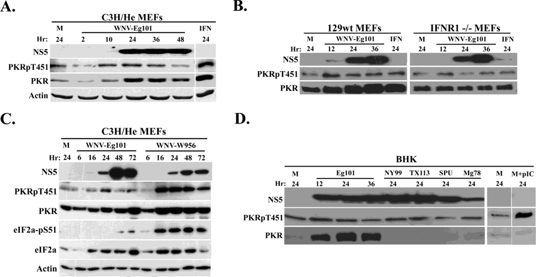
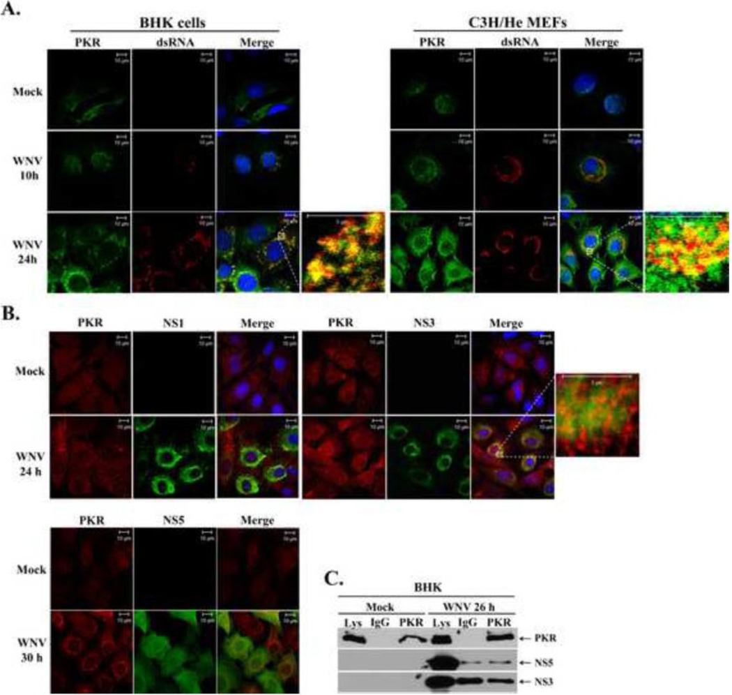
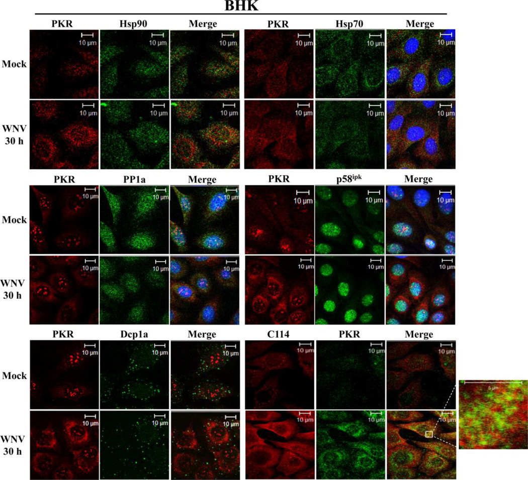
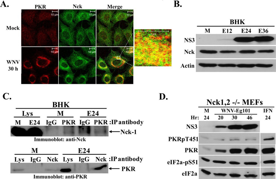
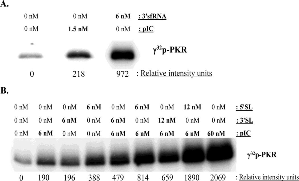
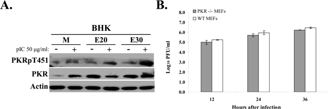
dsRNA-activated protein kinase (PKR) is activated by viral dsRNAs and phosphorylates eIF2a reducing translation of host and viral mRNA. Although infection with a chimeric West Nile virus (WNV) efficiently induced PKR and eIF2a phosphorylation, infections with natural lineage 1 or 2 strains did not. Investigation of the mechanism of suppression showed that among the cellular PKR inhibitor proteins tested, only Nck, known to interact with inactive PKR, colocalized and co-immunoprecipitated with PKR in WNV-infected cells and PKR phosphorylation did not increase in infected Nck1,2-/- cells. Several WNV stem-loop RNAs efficiently activated PKR in vitro but not in infected cells. WNV infection did not interfere with intracellular PKR activation by poly(I:C) and similar virus yields were produced by control and PKR-/- cells. The results indicate that PKR phosphorylation is not actively suppressed in WNV-infected cells but that PKR is not activated by the viral dsRNA in infected cells.


Figures

Figure 1
Figure 1. Analysis of PKR phosphorylation in WNV-infected cells
(A) C3H/He MEFs were mock-infected (M), or infected with WNV Eg101 (MOI of 5) for the indicated times or treated with 100 U/ml universal type I IFN for 24 h (IFN). (B) 129wt or IFNR1−/− MEFs were mock-infected(M) or infected with WNV Eg101 (MOI of 5) for the indicated times or treated with 100 U/ml universal Type I IFN for 24 h.(C) C3H/He MEFs were mock-infected or infected with WNV Eg101 or WNV W956 at a MOI of 1 for the indicated times. (D) BHK cells were mock-infected (M) or infected with WNV Eg101 (MOI of 5) for the indicated times or infected with various strains of WNV (NY99, TX113, SPU and Mg78) for 24 h or transfected with 50 µg/ml poly(IC) for 2 h (pIC). PKR, phopho-Thr451 PKR, eIF2a, phosphor-S51 eIF2a, WNV-NS5 and actin were detected in cell lysates by Western blotting after separation of proteins by 10% SDS-PAGE.
Figure 2
Figure 2. PKR colocalization with sites of WNV replication
(A) Analysis of PKR colocalization with viral replication complexes. BHK cells and MEFs were mock-infected or infected with WNV Eg101 (MOI of 5). At 10 and 24 h after infection, cells were permeabilized, fixed, and blocked overnight. Cells were stained with anti-PKR and anti-dsRNA antibodies and then AlexaFluor488 (green) and AlexaFluor594 (red) conjugated secondary antibodies, respectively. Cell nuclei were stained with Hoechst. (B) Analysis of PKR colocalization with viral nonstructural proteins. BHK cells and MEFs were mock-infected or infected with WNV Eg101 (MOI of 5). At 24 and 30 h after infection, cells were permeabilized, fixed, and blocked overnight. Cells were stained with anti-PKR and either anti-NS5, anti-NS3 or anti-NS1 antibodies and then AlexaFluor488 (green) and AlexaFluor594 (red) conjugated secondary antibodies, respectively. Cell Nuclei were stained with Hoechst. (C) Analysis of PKR interaction with viral NS3 or NS5 proteins in infected cells. BHK cells were mock-infected (M), or infected with WNV Eg101 (MOI of 5). At 26 h after infection, cells were lysed, S2 fractions were prepared and rabbit anti-PKR antibody or a rabbit IgG was used for immunoprecipitation. Immunoprecipitated proteins were separated by 10% SDS-PAGE and detected by Western blotting using anti-NS5 or anti-NS3 antibodies.
Figure 3
Figure 3. Analysis of PKR colocalization with known cellular PKR inhibitors in WNV-infected cells
Colocalization of PKR with known cellular PKR inhibitors. BHK cells were mock-infected or infected with WNV Eg101 (MOI of 5). At 30 h after infection, cells were permeabilized, fixed and blocked overnight. Cells were stained with anti-PKR and an antibody to a cellular PKR inhibitor and then with AlexaFluor488 (green) and AlexaFluor594/555 (red) conjugated secondary antibodies. PKR was detected with a mouse monoclonal anti-PKR antibody in the PP1a, p58ipk and Dcp1a experiments and with a rabbit polyclonal antibody in the Hsp70, Hsp90 and C114 experiments.
Figure 4
Figure 4. Analysis of Nck-PKR interactions in WNV-infected cells
(A) Analysis of colocalization of PKR with Nck. BHK cells were mock-infected or infected with WNV Eg101(MOI of 5). At 30 h after infection, cells were permeabilized, fixed and blocked overnight. Cells were stained with mouse monoclonal anti-PKR and rabbit polyclonal anti-Nck antibodies and then with AlexaFluor488 (green) and AlexaFluor594/555 (red) conjugated secondary antibodies. (B) Analysis of Nck protein levels in WNV-infected BHK cells. Cells were mock-infected (M), or infected with WNV Eg101 (MOI of 5) for the indicated times. NS3, Nck-1, and actin were detected by Western blotting after separation of proteins by 10% SDS-PAGE. (C) Co-immunoprecipitation of Nck and PKR. BHK cells were mock-infected (M) or infected with WNV Eg101 (MOI of 5). At 24 h after infection, cells were lysed and S2 fractions were prepared. Rabbit anti-PKR antibody or a rabbit anti-Nck antibody was used for immunoprecipitation; rabbit IgG was used as a control antibody.. Immunoprecipitated proteins were separated by 10% SDS-PAGE and detected by Western blotting using a rabbit anti-Nck antibody (top panel) or a mouse anti-PKR antibody (bottom panel). (D) Analysis of PKR phosphorylation in WNV-infected Nck-knockout cells. Nck-1,2−/− MEFs were mock-infected (M) or infected with WNV Eg101 (MOI of 5) for the indicated times or treated with 100 U/ml universal type I IFN for 24 h (IFN). NS3, total PKR, phopho-Thr451 PKR, total eIF2a and phospho-Ser51 eIF2a were detected by Western blotting after separation of proteins by 10% SDS-PAGE. Blots shown are representative of at least two independent experiments.
Figure 5
Figure 5. In vitro PKR autophosphorylation assays
(A) Reaction mixtures containing 150 ng of purified PKR alone or with the indicated concentrations of in vitro transcribed WNV 3'sfRNA or poly(I:C) were incubated as described in Materials and Methods. (B) Reaction mixtures containing 150 ng of purified PKR alone or with the indicated concentrations of in vitro transcribed WNV 3'SL or 5'SL RNA or with poly(I:C) were incubated for 30 min as described in Materials and Methods, or preincubated with the 3'SL or 5'SL for 10 min and then with poly(I:C) for 30 min. γ32P-ATP was included in the reactions as a phosphate donor. Image Gauge 3.2 software was used to measure the relative band intensities of images acquired using a Fuji BAS 2500 analyzer. Results are representative of two independent experiments.
Figure 6
Figure 6. Analysis of active suppression of PKR activation in infected cells and of PKR anti-flaviviral activity
(A) Poly(I:C)-mediated PKR autophosphorylation in WNV-infected cells. BHK cells were mock-infected or infected with WNV Eg101 (MOI of 5). At 20 or 30 h after infection, cells were transfected with 50 µg/ml of poly(I:C) in Celfectin II (+) or transfection reagent alone (−) for 1.5 h before cell lysis. PKR, phopho-Thr451 PKR and actin were detected in cell lysates by Western blotting after separation of proteins by 10% SDS-PAGE. (B) Viral yields produced by PKR−/− and wildtype MEFs infected with WNV Eg101 (MOI of 5). Samples of culture fluid were harvested at the indicated times, and infectivity titers were determined by plaque assays done in duplicate on BHK cells. Virus titers are expressed as log10 PFU/ml. Error bars indicate ± standard error of the mean (SEM) (n = 3).

References

    1. Arnaud N, Dabo S, Maillard P, Budkowska A, Kalliampakou KI, Mavromara P, Garcin D, Hugon J, Gatignol A, Akazawa D, Wakita T, Meurs EF. Hepatitis C virus controls interferon production through PKR activation. PLoS One. 2010;5:e10575. - PMC - PubMed
    1. Brinton MA. The molecular biology of West Nile Virus: a new invader of the western hemisphere. Annu Rev Microbiol. 2002;56:371–402. - PubMed
    1. Cardin E, Larose L. Nck-1 interacts with PKR and modulates its activation by dsRNA. Biochem Biophys Res Commun. 2008;377:231–235. - PubMed
    1. Cardin E, Latreille M, Khoury C, Greenwood MT, Larose L. Nck-1 selectively modulates eIF2alphaSer51 phosphorylation by a subset of eIF2alpha-kinases. FEBS J. 2007;274:5865–5875. - PubMed
    1. Conn GL. Expression of active RNA-activated protein kinase (PKR) in bacteria. Biotechniques. 2003;35:682–684. 686. - PubMed

Publication types

MeSH terms

Substances