. 2011 Oct 28;35(4):596-610.
doi: 10.1016/j.immuni.2011.08.001. Epub 2011 Oct 6.

Pivotal role of dermal IL-17-producing γδ T cells in skin inflammation

Affiliations

Pivotal role of dermal IL-17-producing γδ T cells in skin inflammation

Yihua Cai et al. Immunity. .

Erratum in

  • Immunity. 2011 Oct 28;35(4):649

Abstract

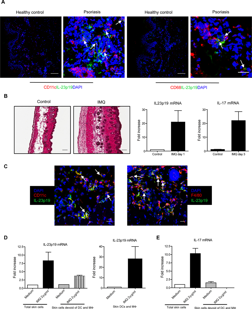
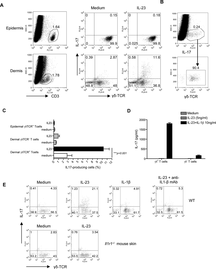
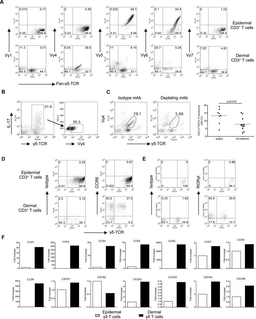
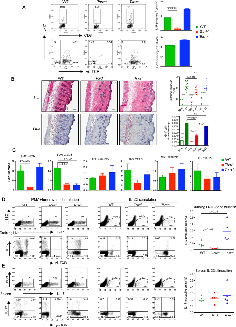
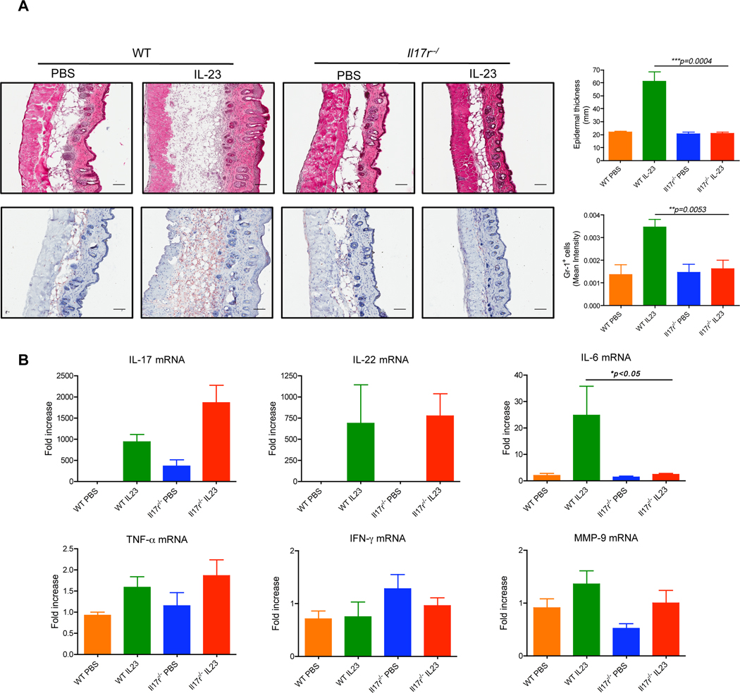
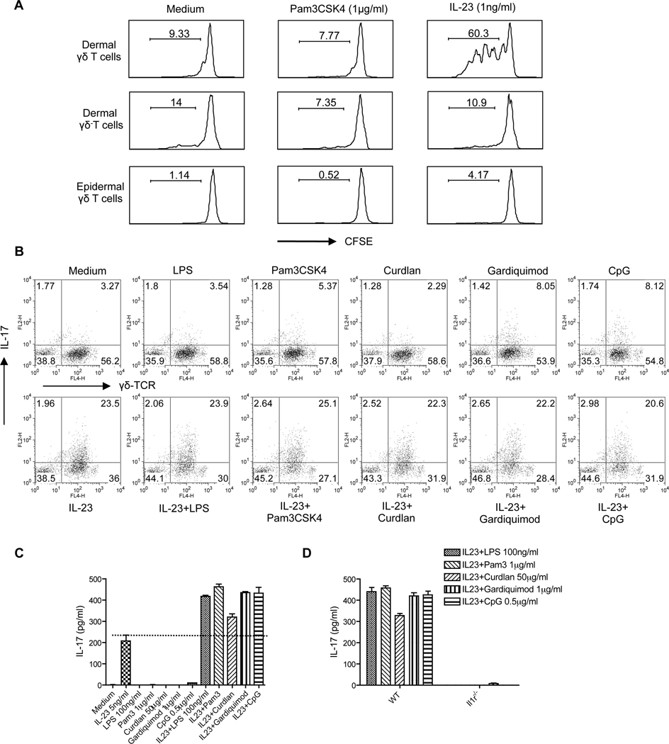
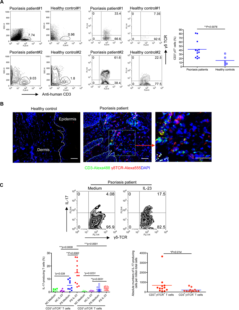
Interleukin-23 (IL-23) and CD4(+) T helper 17 (Th17) cells are thought to be critical in psoriasis pathogenesis. Here, we report that IL-23 predominantly stimulated dermal γδ T cells to produce IL-17 that led to disease progression. Dermal γδ T cells constitutively expressed the IL-23 receptor (IL-23R) and transcriptional factor RORγt. IL-17 production from dermal γδ T cells was independent of αβ T cells. The epidermal hyperplasia and inflammation induced by IL-23 were significantly decreased in T cell receptor δ-deficient (Tcrd(-/-)) and IL-17 receptor-deficient (Il17ra(-/-)) mice but occurred normally in Tcra(-/-) mice. Imiquimod-induced skin pathology was also significantly decreased in Tcrd(-/-) mice. Perhaps further promoting disease progression, IL-23 stimulated dermal γδ T cell expansion. In psoriasis patients, γδ T cells were greatly increased in affected skin and produced large amounts of IL-17. Thus, IL-23-responsive dermal γδ T cells are the major IL-17 producers in the skin and may represent a novel target for the treatment of psoriasis.


Figures

Figure 1
Figure 1. DCs and Mϕ are the major cellular source of IL-23 in psoriatic skin
(A) Frozen skin sections from patients with psoriatic lesions (scale bar, 25µm) and healthy controls (scale bar, 100µm) were stained with anti-human IL-23p19 (green), anti-human CD11c (red) or anti-human CD68 (red) and DAPI (blue) for immunofluorescent staining. (B) Representative H&E-stained sections of the back skin of C57BL/6 WT mice treated for 3 consecutive days with control cream or IMQ are shown (scale bar, 100µm). IL-17 and IL-23p19 mRNA concentrations were measured by qPCR. Data are shown as mean± SEM. (C) Frozen sections from 3 days of IMQ-treated mouse back skin were co-stained with anti-mouse IL-23p19 (green) and anti-mouse CD11c (red), or anti-mouse F4/80 (red) and DAPI (blue) for immunofluorescent staining. Scale bar, 25µm. (D, E) Whole mouse skin cells, skin cells devoid of DCs and Mϕ or purified skin DCs and Mϕ were stimulated with IMQ for 24 hr or 3 hr and IL-23p19 (D) and IL-17 (E) mRNA concentrations were measured by qPCR. Data are shown as mean± SEM.
Figure 2
Figure 2. Dermal γδ T cells are the predominant IL-17 producers upon IL-23 stimulation in the skin
Intracellular IL-17 production assessed by flow cytometry on epidermal and dermal cell suspensions from C57BL/6 WT mice that were stimulated with IL-23 for 18 hr. (A) Cells were gated on CD3+ T cells. (B) Dermal IL-17 producing cells were gated and calculated for γδ TCR expression. (C) Percentages of IL-17-producing cells in dermal CD3+γδ TCR+ cells, CD3+γδ TCR cells and epidermal CD3+γδ TCR+ cells were analyzed from twelve independent experiments. Data are shown as mean± SEM. ***P<0.001 (unpaired Student’s t-test). (D) Dermal CD3+γδ TCR+ cells and CD3+γδ TCR cells were sorted and then stimulated with IL-23 in the presence or absence of IL-1β for 2 days. IL-17 production was measured by ELISA. (E) Skin cells from WT or Il1r1−/− mice were stimulated with IL-23, IL-1β or IL-23 plus IL1β mAb or isotype control mAb. Intracellular IL-17 concentration was assessed by flow cytometry. Cells were gated on CD3+ T cells.
Figure 3
Figure 3. Phenotypic analysis of dermal γδ T cells versus epidermal γδ T cells
(A) Epidermal and dermal cell suspensions were stained with a panel of different Vγ TCR antibodies (Vγ1, Vγ4, Vγ5, Vγ6 and Vγ7) and analyzed by flow cytometry. Flow plots gated on CD3+ cells are representative of two independent experiments with similar results. (B) Dermal cell suspensions were stimulated with IL-23 for 18 hr and analyzed for intracellular IL-17 expression by flow cytometry after staining with different Vγ TCR antibodies. Flow plots gated on CD3+ cells are representative of two independent experiments. (C) Dermal cells from C57BL/6 WT mice receiving mouse Vγ4 mAb or isotype control mAb for three days were stimulated with IL-23 and intracellular IL-17 expression was determined by flow cytometry. Percentages of IL-17+CD3+γδ TCR+ cells were analyzed from three independent experiments. Data are shown as mean ± SEM. *P<0.05 (unpaired Student’s t-test). (D, E) CCR6 (D) and RORγt (E) expression on epidermal and dermal γδ T cells were determined by flow cytometry. (F) Expression of chemokine receptors and IL-23R mRNA measured by qPCR in FACS-sorted epidermal or dermal γδ T cells. The figure shows fold changes of the indicated genes normalized for β-MG mRNA versus the epidermal γδ T cells.
Figure 4
Figure 4. γδ T cells are critical in IL-23-induced skin inflammation and acanthosis
(A) Dermal cell suspensions from C57BL/6 WT, Tcrd−/− and Tcra−/− mice were stimulated with IL-23 and analyzed for intracellular IL-17 expression by flow cytometry. Flow plots gated on CD3+ cells are representative of two independent experiments. (B) C57BL/6 WT (n=12), Tcrd−/− (n=12) and Tcra−/− (n=8) mice received daily intradermal injections with IL-23 or vehicle control for 4 days. Representative H&E-stained sections and frozen sections stained with Gr-1 are shown. Epidermal thickness and Gr-1 infiltration were measured at day 4. Scale bar, 100 µm. Data are combined from two independent experiments. *P<0.05, ***p<0.001; n.s., not significant (unpaired Student’s t-test). (C) IL-17, IL-22, IL-6, TNF-α, MMP-9 and IFN-γ mRNA concentrations were measured by qPCR. The figure shows fold changes normalized for β-MG mRNA versus IL-23-injected skin from Tcrd−/− mice. Data are representative of two independent experiments and shown as mean±SEM. (D, E) Skin draining LN cells (D) or splenocytes (E) from IL-23-treated mice were stimulated with PMA plus ionomycin or IL-23 and intracellular IL-17 expression was determined by flow cytometry. Upper panels are representative dot plots gated on the total cells. The lower panels are representative dot plots gated on the CD3+ T cells. *P<0.05, **P<0.01 (unpaired Student’s t-test).
Figure 5
Figure 5. IL-17R expression is essential for IL-23-induced epidermal hyperplasia
C57BL/6 WT (n=5) and Il17ra−/ − (n=5) mice received daily intradermal injections with IL-23 or vehicle control for 4 days. (A) Representative H&E-stained sections and frozen sections stained with Gr-1 are shown. Epidermal thickness and Gr-1 infiltration were measured at day 4. Scale bar, 100 µm. Data are shown as mean± SEM. (B) IL-17, IL-22, IL-6, TNF-α, MMP-9 and IFN-γ mRNA concentrations were measured by qPCR. The figure shows fold changes normalized for β-MG mRNA versus control skin from WT mice. Data are shown as mean± SEM. **P<0.01, ***P<0.001 (unpaired Student’s t-test).
Figure 6
Figure 6. Dermal γδ T cell in vitro expansion and IL-17 production stimulated by IL-23 combined with specific pathogenic products
(A) Skin cell suspensions were labeled with CFSE and then stimulated with Pam3CSK4 or IL-23 for 3 days. Cells were harvested and stained with CD3 and γδ TCR mAbs. (B) Skin cells were stimulated with IL-23 alone, different pathogen products (LPS, Pam3CSK4, Curdlan, Gardiquimon, or CpG) or IL-23 plus different pathogenic products for 2 days. Intracellular IL-17 production was determined by flow cytometry. Cells were gated on CD3+ T cells. (C) Supernatants harvested from (B) were measured for IL-17 concentration by ELISA. Data are shown as mean± SEM. (D) Skin cells from WT and Il1r1−/− mice were stimulated with IL-23 plus different pathogen products for 2 days. Supernatants were harvested and IL-17 concentrations were determined by ELISA. Data are shown as mean± SEM. Data are representative of at least three independent experiments with similar results.
Figure 7
Figure 7. Skin lesions from psoriasis patients display high frequency of IL-17-secreting γδ T cells
(A) Dermal cells from psoriatic lesions or healthy controls were analyzed for CD3 and γδ TCR expression by flow cytometry. Two donors from each group (both dot plots and contour plots) were shown. Flow plots gated on CD3+ cells are representative from 12 patients and 6 healthy controls. Percentage of CD3+γδ TCR+ cells is shown as mean± SEM. Statistical analysis was performed by using a two-tailed Mann Whitney test. **p<0.01. (B) Frozen sections from psoriatic lesion and healthy controls were stained with human γδ TCR mAb (red), CD3 mAb (green) and DAPI (blue) for immuno-fluorescent staining. Scale bar, 10 µm. (C) Dermal cell suspensions from psoriatic lesion (PS) and healthy control (NC) were stimulated with IL-23 and IL-17 expression was determined by flow cytometry. Cells were gated on CD3+γδ TCR+ or CD3+γδ TCR cells. Percentage of IL-17+CD3+γδ TCR+ and IL-17+CD3+γδ TCR cells is shown as mean ± SEM. Absolute numbers of IL-17-producing cells per million cells are shown from 12 patients. *p<0.05, **p<0.01, ***p<0.001 (a Mann Whitney test).

Comment in

  • Inflammation: Under the skin.
    Papatriantafyllou M. Papatriantafyllou M. Nat Rev Immunol. 2011 Nov 4;11(12):800. doi: 10.1038/nri3113. Nat Rev Immunol. 2011. PMID: 22051889 No abstract available.

References

    1. Aliahmadi E, Gramlich R, Grutzkau A, Hitzler M, Kruger M, Baumgrass R, Schreiner M, Wittig B, Wanner R, Peiser M. TLR2-activated human langerhans cells promote Th17 polarization via IL-1beta, TGF-beta and IL-23. Eur J Immunol. 2009;39:1221–1230. - PubMed
    1. Blauvelt A. T-helper 17 cells in psoriatic plaques and additional genetic links between IL-23 and psoriasis. J Invest Dermatol. 2008;128:1064–1067. - PMC - PubMed
    1. Boniface K, Guignouard E, Pedretti N, Garcia M, Delwail A, Bernard FX, Nau F, Guillet G, Dagregorio G, Yssel H, et al. A role for T cell-derived interleukin 22 in psoriatic skin inflammation. Clin Exp Immunol. 2007;150:407–415. - PMC - PubMed
    1. Born WK, Yin Z, Hahn YS, Sun D, O'Brien RL. Analysis of gammadelta T cell functions in the mouse. J Immunol. 2010;184:4055–4061. - PMC - PubMed
    1. Buonocore S, Ahern PP, Uhlig HH, Ivanov II, Littman DR, Maloy KJ, Powrie F. Innate lymphoid cells drive interleukin-23-dependent innate intestinal pathology. Nature. 2010;464:1371–1375. - PMC - PubMed

Publication types

MeSH terms