

Comment
doi: 10.1016/j.cmet.2011.09.002.
DisseCCTing phospholipid function in lipid droplet dynamics
Affiliations
- PMID: 21982702
- PMCID: PMC3211137
- DOI: 10.1016/j.cmet.2011.09.002
Item in Clipboard
Comment
DisseCCTing phospholipid function in lipid droplet dynamics
Cell Metab.
.
Abstract
Phospholipids provide an amphipathic barrier between lipid droplets and the cytoplasm of cells. In this issue of Cell Metabolism, Krahmer and colleagues (2011) define a role for phosphatidylcholine in preventing lipid droplet coalescence and show that the rate-limiting enzyme in phosphatidylcholine synthesis is activated through binding to lipid droplets.
Copyright © 2011 Elsevier Inc. All rights reserved.
Figures
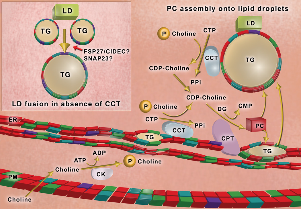
Dietary choline is taken up across the plasma membrane (PM) and is phosphorylated by choline kinase (CK). Phosphocholine is converted to CDP-choline by CTP:phosphocholine cytidylyltransferase (CCT), which binds to endoplasmic reticulum (ER) membranes and the phospholipid monolayer (colored blocks) of lipid droplets (LD) via an amphipathic helical domain (Krahmer, et al, 2011); membrane binding activates the enzyme. CDP-choline is condensed with diglyceride (DG) to make phosphatidylcholine (PC) by an integral protein of the ER membrane, CDP-choline:1,2 diacylglycerol cholinephosphotransferase (CPT). Phosphatidylcholine (represented by red blocks) is the most abundant phospholipid in cellular membranes and the phospholipid monolayer of lipid droplets. Lipid droplets are thought to originate as a lens of triglyceride (TG) within the membrane bilayer of the ER. Following expansion of the triglyceride core, lipid droplets bud out of the ER to become independent organelles in the cytoplasm of the cell. When phosphatidylcholine is limiting in the phospholipid monolayer coating the lipid droplet (Inset), adjacent lipid droplets coalesce into larger lipid droplets (Krahmer, et al., 2011). Lipid droplet-associated proteins may also promote lipid droplet fusion; candidates include SNAP23, and Fsp27/CIDEC. (Illustration by R. Hasney)
Comment on
-
Phosphatidylcholine synthesis for lipid droplet expansion is mediated by localized activation of CTP:phosphocholine cytidylyltransferase.Cell Metab. 2011 Oct 5;14(4):504-15. doi: 10.1016/j.cmet.2011.07.013. Cell Metab. 2011. PMID: 21982710 Free PMC article.
References
-
- Bartz R, Li W-H, Venables B, Zehmer JK, Roth MR, Welti R, Anderson RGW, Liu P, Chapman KD. J. Lipid Res. 2007;48:837–847. - PubMed
-
- Bostrom P, Andersson L, Rutberg M, Perman J, Lidberg U, Johansson BR, Fernandez-Rodriguez J, Ericson J, Nilsson T, Boren J, Olofsson S-O. Nature Cell Biol. 2007;9:1286–1293. - PubMed
Publication types
Grants and funding
LinkOut - more resources
Full Text Sources
