Review
. 2012 Feb;24(2):378-387.
doi: 10.1016/j.cellsig.2011.09.028. Epub 2011 Oct 1.

The renaissance of Ca2+-binding proteins in the nervous system: secretagogin takes center stage

Affiliations
Review

The renaissance of Ca2+-binding proteins in the nervous system: secretagogin takes center stage

Alán Alpár et al. Cell Signal. 2012 Feb.

Abstract

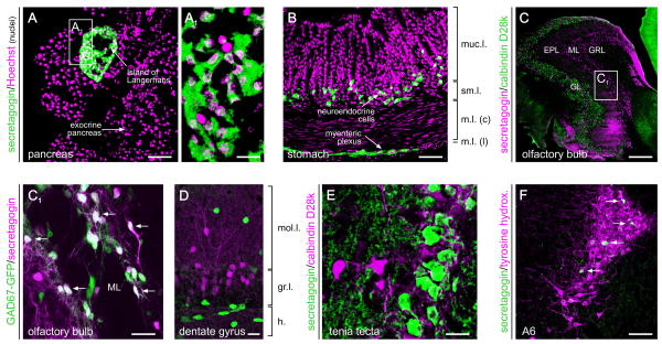
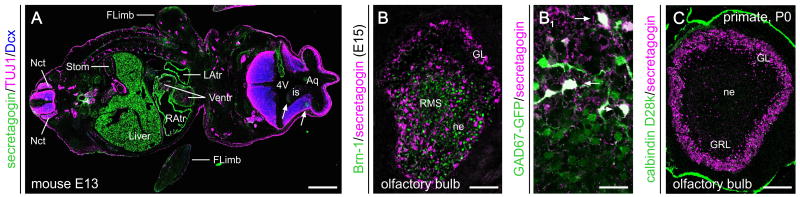
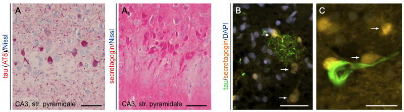
Effective control of the Ca(2+) homeostasis in any living cell is paramount to coordinate some of the most essential physiological processes, including cell division, morphological differentiation, and intercellular communication. Therefore, effective homeostatic mechanisms have evolved to maintain the intracellular Ca(2+) concentration at physiologically adequate levels, as well as to regulate the spatial and temporal dynamics of Ca(2+)signaling at subcellular resolution. Members of the superfamily of EF-hand Ca(2+)-binding proteins are effective to either attenuate intracellular Ca(2+) transients as stochiometric buffers or function as Ca(2+) sensors whose conformational change upon Ca(2+) binding triggers protein-protein interactions, leading to cell state-specific intracellular signaling events. In the central nervous system, some EF-hand Ca(2+)-binding proteins are restricted to specific subtypes of neurons or glia, with their expression under developmental and/or metabolic control. Therefore, Ca(2+)-binding proteins are widely used as molecular markers of cell identity whilst also predicting excitability and neurotransmitter release profiles in response to electrical stimuli. Secretagogin is a novel member of the group of EF-hand Ca(2+)-binding proteins whose expression precedes that of many other Ca(2+)-binding proteins in postmitotic, migratory neurons in the embryonic nervous system. Secretagogin expression persists during neurogenesis in the adult brain, yet becomes confined to regionalized subsets of differentiated neurons in the adult central and peripheral nervous and neuroendocrine systems. Secretagogin may be implicated in the control of neuronal turnover and differentiation, particularly since it is re-expressed in neoplastic brain and endocrine tumors and modulates cell proliferation in vitro. Alternatively, and since secretagogin can bind to SNARE proteins, it might function as a Ca(2+) sensor/coincidence detector modulating vesicular exocytosis of neurotransmitters, neuropeptides or hormones. Thus, secretagogin emerges as a functionally multifaceted Ca(2+)-binding protein whose molecular characterization can unravel a new and fundamental dimension of Ca(2+)signaling under physiological and disease conditions in the nervous system and beyond.


Conflict of interest statement

Conflict of interest statement: The Authors declare no conflict of interest.

Figures

Fig. 1
Fig. 1. Organ systems distribution of secretagogin during adulthood
(A, A1) Secretagogin is expressed at particularly high levels by pancreatic β cells in the islands of Langerhans. Neuroendocrine cells, such as those in the principal part of gastric glands (B), also harbour secretagogin. In the nervous system, secretagogin is expressed by interneurons in the periglomerular and plexiform layers of the olfactory bulb (C, C1), where it can co-exist with glutamic acid decarboxylase (GAD, arrows), the enzyme synthesizing GABA. Secretagogin has also been found in granular cells of the dentate gyrus (D) and tenia tecta (E), as well as the locus coeruleus (F) containing noradrenergic cells (group A6). Arrows in (F) identify secretagogin+ cells intermingled with tyrosine hydroxylase-expressing neurons. Abbreviations: EPL, external plexiform layer; GL, glomerular layer; GRL, granular layer; ML, mitral layer; m.l. (c/l), muscular layer (central/lateral); muc.l., mucosal layer; sm.l., submucosal layer. Scale bar = 300 μm (C), 70 μm (A,B,F), 20 μm (A1,C1,D,E).
Fig. 2
Fig. 2. Secretagogin expression during foetal development
(A) Secretagogin immunoreactivity in the central nervous system (arrows) and at the periphery (e.g., heart, liver, stomach, gonads) of a mouse embryo at embryonic day (E) 13. (B) Secretagogin+ neurons populate the olfactory bulb by E15, and differentiate into GABAergic neurons as revealed in GAD67-EGFP reporter mice (B1). (C) Secretagogin expression is confined predominantly to periglomerular neurons in the olfactory bulb of grey mouse lemur (Microcebus murinus, Primates) by birth. Abbreviations: 4V, fourth ventricle; Aq, aqueduct; FLimb, forelimb; is, isthmus; GL, glomerular layer; GRL, granular layer of the olfactory bulb; L/RAtr, left/right atrium of the heart; Nct, neural crest; ne, neuroepithel; RMS, rostral migratory stream; Stom, stomach; Ventr, ventricle of the heart. Scale bar = 500 μm (A), 200 μm (C), 70 μm (B), 20 μm (B1).
Fig. 3
Fig. 3. Secretagogin distribution in Alzheimer's disease
(A) Hyperphosphorylated tau immunoreactivity (AT8 staining, in red) in the CA3 subfield of the human hippocampus in Alzheimer's. (A1) Secretagogin immunoreactivity in a consecutive (adjacent) section decorates neurons whose distribution is complementary to AT8+ cells in (A). Nissl stain was used as counterstain (in blue). (B) Secretagogin+/AT8-neurons (arrows) in the vicinity of AT8+ dystrophic neurites accumulating in a putative neuritic plaque. Note the mutually exclusive patterns of immunoreactivity. (C) High-power image of a hyperphosphorylated tau-laden neuron surrounded by secretagogin+ cells (arrows). Scale bar = 100 μm (AA1). 50 μm (B), 20 μm (C).

References

    1. Clapham DE. Cell. 2007;131:1047–1058. - PubMed
    1. Burnashev N, Rozov A. Cell Calcium. 2005;37:489–495. - PubMed
    1. Pang ZP, Sudhof TC. Curr Opin Cell Biol. 2010;22:496–505. - PMC - PubMed
    1. Lipton SA, Nicotera P. Cell Calcium. 1998;23:165–171. - PubMed
    1. Lipton SA, Rosenberg PA. N Engl J Med. 1994;330:613–622. - PubMed

Publication types

MeSH terms

LinkOut - more resources