Review
. 2011 Oct;22(8):858-64.
doi: 10.1016/j.semcdb.2011.09.013. Epub 2011 Oct 1.

Translating cell polarity into tissue elongation

Affiliations
Review

Translating cell polarity into tissue elongation

Athea Vichas et al. Semin Cell Dev Biol. 2011 Oct.

Abstract

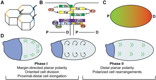
Planar cell polarity, the orientation of single-cell asymmetries within the plane of a multicellular tissue, is essential to generating the shape and dimensions of organs and organisms. Planar polarity systems align cell behavior with the body axes and orient the cellular processes that lead to tissue elongation. Using Drosophila as a model system, significant progress has been made toward understanding how planar polarity is generated by biochemical and mechanical signals. Recent studies using time-lapse imaging reveal that cells engage in a number of active behaviors whose orientation and dynamics translate planar cell polarity into tissue elongation. Here we review recent progress in understanding the cellular mechanisms that link planar polarity to large-scale changes in tissue structure.


Figures

Fig. 1
Fig. 1
Planar cell polarity and tissue elongation in the Drosophila wing. (A) The core planar cell polarity (PCP) proteins localize to proximal (orange) and distal (blue) cell surfaces and are required for planar polarized wing hair formation. (B) Four-jointed (Fj) phosphorylates Fat and Dachsous (Ds) and regulates their interaction. The core PCP proteins form molecularly distinct proximal (P) and distal (D) complexes. (C) Graded expression of Ds (green) and Fj (red) may provide spatial information important for planar polarity. (D) Proximal–distal planar polarity develops from an initially margin-directed pattern (indicated by green arrowheads) over a 32-h period during pupal development. In Phase I (early pupal stages), contraction of the proximal wing hinge (blue) is proposed to generate a force that reorients planar polarity (gray arrows), cell rearrangements, cell divisions, and cell elongation to align with the proximal–distal axis, causing the wing blade to narrow and lengthen. In Phase II (mid-pupal stages), new contacts between cells are assembled parallel to the proximal–distal axis, increasing hexagonal packing and the alignment of planar polarity. Diego (Dgo), Dishevelled (Dsh), Four-jointed (Fj), Flamingo (Fmi), Frizzled (Fz), Prickle (Pk), Strabismus (Stbm). (For interpretation of the references to color in this figure legend, the reader is referred to the web version of the article.)
Fig. 2
Fig. 2
Axis elongation in the Drosophila embryo. (A) During axis elongation, the germband epithelium (dark gray) lengthens 2.5-fold along the anterior–posterior (AP) axis and narrow along the dorsal–ventral (DV) axis. 80% of this elongation occurs in the first 30–40 min and is driven primarily by cell rearrangement. (B) During neighbor exchange, a single myosin-positive cell–cell interface (red) contracts, forming a 4-cell vertex that resolves through the formation of a new interface (blue) between a dorsal and ventral cell. (C) During rosette formation, the coordinated contraction of several consecutive myosin-positive interfaces (red) generates a multicellular rosette structure that resolves in a perpendicular direction, promoting elongation. Cells are labeled with E-cadherin: GFP. Anterior left, ventral down. Scale bar = 10 μm. (For interpretation of the references to color in this figure legend, the reader is referred to the web version of the article.)
Fig. 3
Fig. 3
Elongation of the Drosophila egg chamber. (A) During egg chamber elongation, the Drosophila egg undergoes a 2.5-fold increase in aspect ratio in a process involving multiple rotations (blue arrow) of the entire egg chamber around its long axis in stages 5–9 (~20 h) and periodic contractions of a planar polarized basal actomyosin network in stages 9–10 (~16 h) that produce transient oscillations in basal area during a period of rapid egg chamber growth. (B) During rotation, actin filaments at the basal surface of the follicle cells (red) and collagen IV fibrils in the ECM (green) become aligned parallel to the DV axis. Anterior left, ventral down. (For interpretation of the references to color in this figure legend, the reader is referred to the web version of the article.)

References

    1. Zallen JA. Planar polarity and tissue morphogenesis. Cell. 2007;129:1051–63. - PubMed
    1. Skoglund P, Keller R. Integration of planar cell polarity and ECM signaling in elongation of the vertebrate body plan. Curr Opin Cell Biol. 2010;22:589–96. - PMC - PubMed
    1. Gray RS, Roszko I, Solnica-Krezel L. Planar cell polarity: coordinating morphogenetic cell behaviors with embryonic polarity. Dev Cell. 2011;21:120–33. - PMC - PubMed
    1. Axelrod JD. Unipolar membrane association of Dishevelled mediates Frizzled planar cell polarity signaling. Genes Dev. 2001;15:1182–7. - PMC - PubMed
    1. Strutt DI. Asymmetric localization of frizzled and the establishment of cell polarity in the Drosophila wing. Mol Cell. 2001;7:367–75. - PubMed

Publication types

MeSH terms

LinkOut - more resources