Review
. 2012 May 1;64(6):571-88.
doi: 10.1016/j.addr.2011.09.010. Epub 2011 Sep 29.

Transepithelial transport and toxicity of PAMAM dendrimers: implications for oral drug delivery

Affiliations
Review

Transepithelial transport and toxicity of PAMAM dendrimers: implications for oral drug delivery

S Sadekar et al. Adv Drug Deliv Rev. .

Abstract

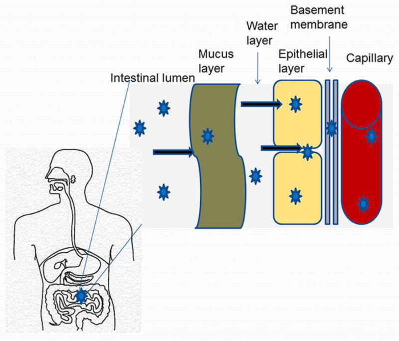
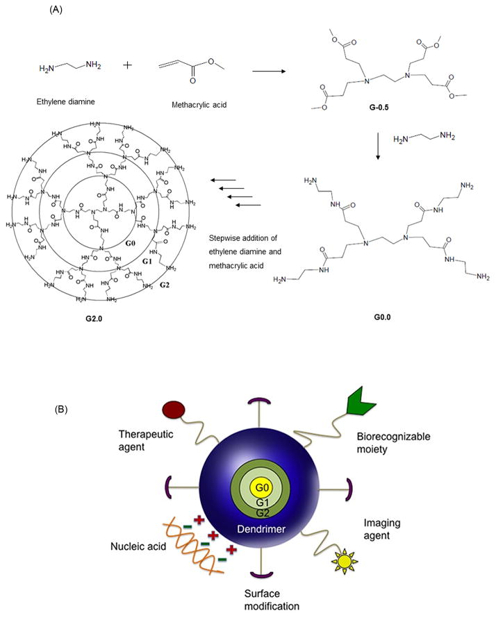
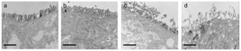
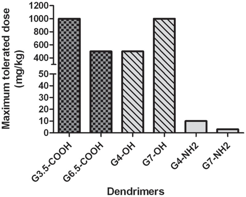
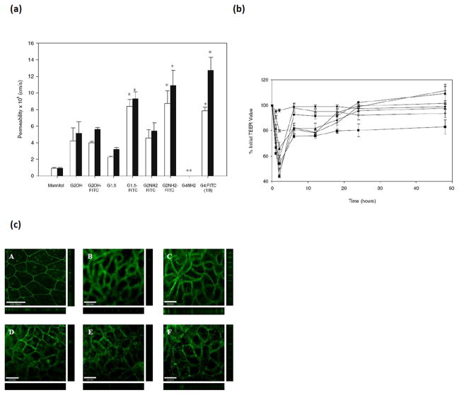
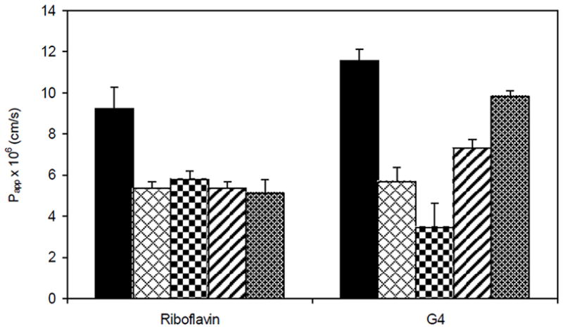
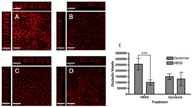
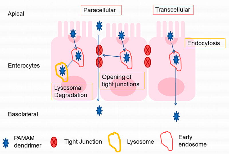
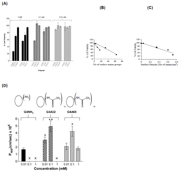
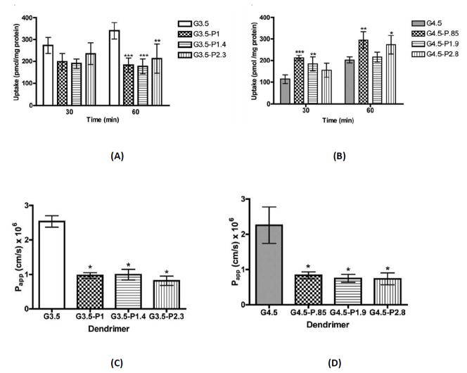
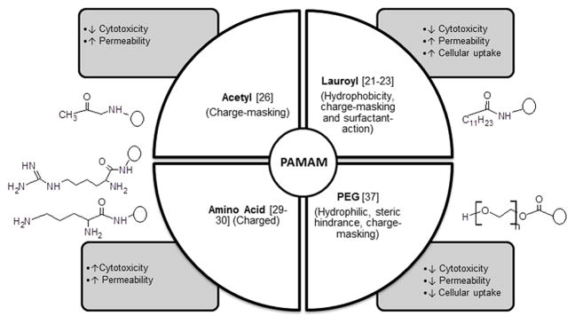
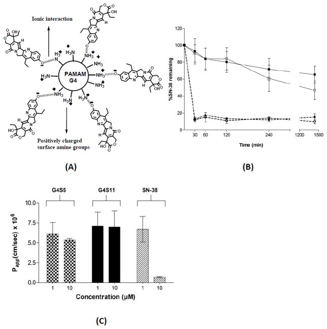
This article summarizes efforts to evaluate poly(amido amine) (PAMAM) dendrimers as carriers for oral drug delivery. Specifically, the effect of PAMAM generation, surface charge and surface modification on toxicity, cellular uptake and transepithelial transport is discussed. Studies on Caco-2 monolayers, as models of intestinal epithelial barrier, show that by engineering surface chemistry of PAMAM dendrimers, it is possible to minimize toxicity while maximizing transepithelial transport. It has been demonstrated that PAMAM dendrimers are transported by a combination of paracellular and transcellular routes. Depending on surface chemistry, PAMAM dendrimers can open the tight junctions of epithelial barriers. This tight junction opening is in part mediated by internalization of the dendrimers. Transcellular transport of PAMAM dendrimers is mediated by a variety of endocytic mechanisms. Attachment or complexation of cytotoxic agents to PAMAM dendrimers enhances the transport of such drugs across epithelial barriers. A remaining challenge is the design and development of linker chemistries that are stable in the gastrointestinal tract (GIT) and the blood stream, but amenable to cleavage at the target site of action. Recent efforts have focused on the use of PAMAM dendrimers as penetration enhancers. Detailed in vivo oral bioavailability of PAMAM dendrimer-drug conjugates, as a function of physicochemical properties will further need to be assessed.


Figures

Figure 1
Figure 1
Gastrointestinal barriers to the oral delivery of polymer therapeutics
Figure 2
Figure 2
Schematics of PAMAM dendrimers for drug delivery; A. Stepwise addition of ethylene diamine and methacrylic acid to alternatively form a half generation, carboxylic acid PAMAM and a full generation amine-terminated PAMAM generation 2. B. A cartoon representing a dendrimer-based delivery system functionalized with therapeutic agent, biorecognizable moiety, imaging agent, nucleic acid and surface-modifying groups. Ref [58], permission to be obtained.
Figure 3
Figure 3
TEM images of Caco-2 cell monolayers after treatment with G4.0-NH2 dendrimers for 2 h: A. control cells; B. 0.01 mM G4.0-NH2; C. 0.1 mM G4.0-NH2; D. 1.0 mM G4NH2 (magnification=12,500x). Scale bars = 1 mm. With permission from Ref [18].
Figure 4
Figure 4
Maximum Tolerated Doses (MTD) in mg/Kg of intravenously administered PAMAM dendrimers to CD-1 mice (n=5). Ref [45], permission to be obtained.
Figure 5
Figure 5
Transepithelial permeability of 14C-mannitol and tight junction modulation in presence of PAMAM dendrimers; (a). The permeability of 14C-mannitol (3.3 μM) in the presence of fluorescently labeled and unlabeled PAMAM dendrimers of donor concentration 1.0 mM across Caco-2 cell monolayers at incubation times of (□) 60 min and (■) 120 min. Permeability values are not reported (**) for dendrimers that cause toxicity. Results are reported as mean +/− standard error of the mean (n = 9). (*) Denotes a significant increase in permeability compared to control (P < 0.001). (b). Transepithelial electrical resistance of Caco-2 cells in the presence of fluorescently labeled PAMAM dendrimers as a function of time: (◆) HBSS transport medium, (■) G2.0-NH2, (▲) G2.0-OH, (×) G1.5-COOH, (+) G2.5-COOH, (●) G3.5-COOH, (◇) G4NH2-FITC (1:8). Results are reported as mean +/− standard error of the mean (n = 9). (c). Staining of the tight junction protein occludin. (A) Caco-2 cells with no polymer treatment. Caco-2 cells incubated for 120 min with 1.0 mM: (B) G2.0-NH2; (C) G2.0-OH, (D) G1.5-COOH; (E) G2.5-COOH; (F) G3.5-COOH. Main panels illustrate the xy plane; horizontal bars illustrate the xz plane; vertical bars illustrate the yz plane. Scale bars equal 100.00 μm. With permission from Ref [15].
Figure 6
Figure 6
Reduced apparent permeability (Papp × 106 cm/s) of Riboflavin (500 nM) and G4.0-NH2 (1 μM) across Caco-2 cell monolayers in the presence of endocytosis inhibitors: Bars from left to right indicate: 1–5 μM brefeldin A; 10 μM colchicine; 1 μg/ml filipin; 200 mM sucrose. Results are reported as mean ± SD (n = 3). **, p < 0.01: ***, p < 0.001. With permission from Ref [22]
Figure 7
Figure 7
Transpeithelial transport and cellular uptake mechanism of PAMAM G3.5-COOH; A–D. Occludin staining in the presence and absence of Oregon green labeled G3.5-COOH dendrimers in Caco-2 cells treated with HBSS or Dynasore. A. G3.5-COOH/HBSS, B. HBSS only C. G3.5-COOH/Dynasore and D. Dynasore only. Main panels illustrate the xy plane; horizontal bars illustrate the xz plane; vertical bars illustrate the yz plane. Scale bars equal 21 μm. E. Quantification of Occludin staining. Results are reported as mean +/− standard deviation with n=4. (***) indicates p<0.001. With permission from Ref [27].
Figure 8
Figure 8
Transepithelial transport mechanisms of PAMAM dendrimers (adapted from Ref [27]).
Figure 9
Figure 9
Cytotoxicity and transepithelial transport of acetylated PAMAM G4.0-NH2; (A) In vitro cell viability of Caco-2 cells after incubation with PAMAM dendrimers (1 mM) for 3 hours. Results are reported as mean +/−SEM (n = 9) where G4NH2, G4A32, G4A60: PAMAM G4.0 with 0, 32 and 60 amine groups acetylated respectively; (B) Relationship between cell viability and number of surface amine groups for (■) G4.0-NH2 (R2 = 0.99) and (▲) G2.0-NH2 analogues (R2 = 1); (C). Relationship between cell viability and surface density (R2 = 0.93); (D) Permeability of G4.0-NH2, G4A32, and G4A60 across Caco-2 cell monolayers after 120 min. Results are reported as mean +/− SEM (n=6). (*) (p < 0.05) and (**) (p < 0.01) denote a significant difference in permeability when compared to permeability of unmodified PAMAM dendrimer at 0.01 mM. (×) Permeability is not evaluated due to cytotoxicity. With permission from Ref [16].
Figure 10
Figure 10
Cellular uptake and transepithelial transport of PAMAM G3.4 and G4.5; Uptake of G3.5 (A) and G4.5 (B) native and differentially PEGylated dendrimers at 0.02 mM for 30 and 60 minutes in Caco-2 cells. n=3, Mean ± standard deviation. (*), (**) and (***) denote significant differences from unmodified dendrimers with p<0.05, p<0.01 and p<0.001 respectively. Apparent permeability of G3.5 (C) and G4.5 (D) native and differentially PEGylated dendrimers at 0.1 mM after a 2 hour incubation time with Caco-2 cell monolayers. n=3, Mean ± standard deviation. (*) denotes a significant difference from unmodified dendrimers with p<0.001. With permission from Ref [24].
Figure 11
Figure 11
Effect of surface modification on PAMAM cytotoxicity, transepithelial permeability and cellular uptake.
Figure 12
Figure 12
Gastrointestinal stability and transpeithelial transport of PAMAM-SN38 complex. A. Schematic representation of G4S5 complex. B. Stability of polymer-SN-38 complexes G4S5 (open squares, solid line) and G4S11 (filled squares, solid line) at pH 7.4, and G4S5 (open circles, dotted line) and G4S11 (filled circles, dotted line) at pH 5. G4S5, G4S11: 5 and 11 moles of SN38 complexed to PAMAM G4.0 respectively. C. Permeability of G4S5, G4S11 and SN-38, across Caco-2 cell monolayers after 120 min With permission from Ref [21].
Figure 13
Figure 13
Synthetic Schemes of G3.5-Gly-SN38 (top) and G3.5-βAla-SN38 (bottom) conjugates. With permission from Ref [28, 53]; Table: Characteristics of G3.5 PAMAM-SN38 conjugates. With permission from Ref [53]
Figure 14
Figure 14
Nuclear fragmentation in HCT-116 cells treated with drug/conjugates. Untreated cells (column 1); 5 nM SN38 (column 2); 40 nM G3.5-gly-SN38 (column 3); 120 nM G3.5-βala-SN38 (column 4). Scale bar is 10 μm. Arrows indicate nuclear fragments. From bottom: 1st row, differential interference contrast image; 2nd row, fluorescence image; 3rd row, overlay of differential interference contrast and fluorescence images. With permission from Ref [53].
Figure 15
Figure 15
Stability and transepithelial transport of G3.5-Gly-SN38 and G3.5-βAla-SN38 conjugates. Release of SN38 was monitored in simulated conditions of the stomach for 2 hours (A), intestine for 24 hours (B) and liver for 48 hours (C). Mean ± standard deviation (n=2). G3.5-Gly-SN38 is represented by red squares and G3.5-βAla-SN38 is represented by purple circles. Buffers without enzymes are depicted as dashed lines with open symbols and buffers with enzymes are depicted as solid lines with filled symbols. (D) Equivalent SN38 flux across differentiated Caco-2 monolayers treated with G3.5-SN38 conjugates and SN38. Equivalent SN38 flux was calculated by multiplying the measured molar flux of the conjugates with the number of SN38 molecules per dendrimer. Mean ± standard deviation (n=4). (***) indicates a significant difference with p<0.001. With permission from Ref [28].

References

    1. Duncan R. Polymer conjugates as anticancer nanomedicines. Nat Rev Cancer. 2006;6:688–701. - PubMed
    1. Duncan R. The dawning era of polymer therapeutics. Nat Rev Drug Discovery. 2003;2:347–360. - PubMed
    1. Hillery A, Lloyd AW, Swarbrick J. Drug delivery and targeting. CRC Press; 2003.
    1. Ponchel G, Irache JM. Specific and non-specific bioadhesive particulate systems for oral delivery to the gastrointestinal tract. Adv Drug Delivery Rev. 1998;34:191–219. - PubMed
    1. Florence AT, Sakthivel T, Toth I. Oral uptake and translocation of a polylysine dendrimer with a lipid surface. J Controlled Release. 2000;65:253–259. - PubMed

Publication types