Review
. 2011 Jul-Sep;7(3):165-79.
doi: 10.4161/org.7.3.18143. Epub 2011 Jul 1.

Planar cell polarity in Drosophila

Affiliations
Review

Planar cell polarity in Drosophila

Saw Myat Thanda W Maung et al. Organogenesis. 2011 Jul-Sep.

Abstract

In all multicellular organisms, epithelial cells are not only polarized along the apical-basal axis, but also within the epithelial plane, giving cells a sense of direction. Planar cell polarity (PCP) signaling regulates establishment of polarity within the plane of an epithelium. The outcomes of PCP signaling are diverse and include the determination of cell fates, the generation of asymmetric but highly aligned structures, such as the stereocilia in the human inner ear or the hairs on a fly wing, or the directional migration of cells during convergence and extension during vertebrate gastrulation. In humans, aberrant PCP signaling can result in severe developmental defects, such as open neural tubes (spina bifida), and can cause cystic kidneys. In this review, we discuss the basic mechanism and more recent findings of PCP signaling focusing on Drosophila melanogaster, the model organism in which most key PCP components were initially identified.
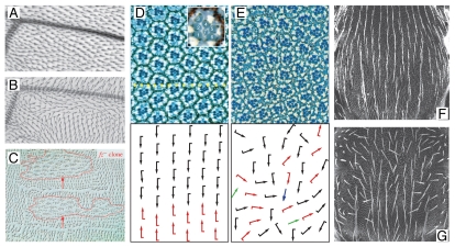
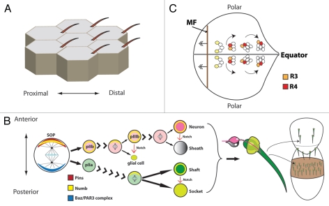
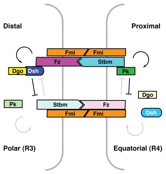
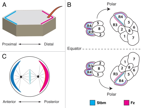

Figures

Figure 1
Figure 1
PCP phenotypes in Drosophila. (A and B) Enlarged view of part of an adult wild type (A) and stbm mutant (B) wing. Note the irregular wing hair swirls of the stbm mutant. Distal is to the left; anterior is up. (C) Trichomes on the cuticle of abdominal tergites. Note the posterior non-autonomy of fz mutant clones (outlined by red lines), where trichomes point anteriorly toward the clone (red arrows) instead of posteriorly. Image courtesy of P. Lawrence (adapted with permission). (D and E) Examples of wild type (D) and fz mutant (E) adult eye sections showing the semi-crystalline arrangement of ommatidia. The schemes below the sections indicate the polarity of ommatidia, with arrows drawn from R3 to R1 and the flag pointing toward R4. Note the randomized chirality and degree of rotation in the fz mutant. Yellow dots represent the equator. The insert in (D) is a high magnification of a single ommatidium with the photoreceptors numbered (R8 is below R7 and thus cannot be seen). (E and F) Scanning electron micrographs of the dorsal thorax of adult flies. (E) In wild type, thoracic bristles are well aligned and point toward the posterior. However, in a fz mutant (F), bristles are misaligned due to the random planar division axis of SOP cells. Images courtesy of P. Adler (adapted with permission).
Figure 2
Figure 2
Schematic of PCP in the fly. (A) Proximal-distal orientation of hairs in a group of adult wing cells. (B) Asymmetric division and spindle orientation of SOPs is determined by anterior localization of Pins and Numb and posterior localization of the Baz/Par3 complex to produce N signaling asymmetries and antero-posterior alignment of the adult bristle. (C) Schematic of a developing third instar eye imaginal disc with the dorso-ventral midline (equator) in gray and the morphogenetic furrow (MF) in brown. The R3/4 pair is initially equivalent (pale yellow). The cell of the R3/4 pair closer to the equator is specified as R3 (yellow) upon Fz-PCP signaling, while its neighbor becomes R4 (red). Ommatidia rotate 90° in opposing directions on either half of the eye.
Figure 3
Figure 3
Model representing asymmetric protein-protein interactions across distal-proximal cell and polar-equatorial cell-cell contacts. See text for details.
Figure 4
Figure 4
Asymmetric localization of core PCP proteins. (A) Fz (pink) and Stbm (blue) are transiently asymmetrically localized in wing cells with Fz localizing to the distal apical membrane and Stbm localizing proximally. (B) In the five-cell precluster of the eye imaginal disc, expression of Fz and Stbm are initially equivalent in the presumptive R3/R4 cells but as development continues, the ommatidium starts to rotate and Fz becomes localized to the R3 side of the R3/R4 membrane border while Stbm localizes to the R4 cell membrane. (C) In the SOP of the bristle Fz localizes to the posterior membrane and Stbm localizes to the anterior membrane before asymmetric division. Where tested, Dsh and Dgo colocalize with Fz, while Pk colocalizes with Stbm (for details see text).

References

    1. Eaton S, Simons K. Apical, basal and lateral cues for epithelial polarization. Cell. 1995;82:5–8. doi: 10.1016/0092-8674(95)90045-4. - DOI - PubMed
    1. Wodarz A, Nathke I. Cell polarity in development and cancer. Nat Cell Biol. 2007;9:1016–1024. doi: 10.1038/ncb433. - DOI - PubMed
    1. Lecuit T, Lenne PF. Cell surface mechanics and the control of cell shape, tissue patterns and morphogenesis. Nat Rev Mol Cell Biol. 2007;8:633–644. doi: 10.1038/nrm2222. - DOI - PubMed
    1. Zallen JA, Blankenship JT. Multicellular dynamics during epithelial elongation. Semin Cell Dev Biol. 2008;19:263–270. doi: 10.1016/j.semcdb.2008.01.005. - DOI - PMC - PubMed
    1. Henderson DJ, Chaudhry B. Getting to the heart of planar cell polarity signaling. Birth Defects Res A Clin Mol Teratol. 2011;91:460–467. doi: 10.1002/bdra.20792. - DOI - PubMed

Publication types

Substances

LinkOut - more resources