

B cells in multiple sclerosis: connecting the dots
- PMID: 21983151
- PMCID: PMC4188435
- DOI: 10.1016/j.coi.2011.09.003
B cells in multiple sclerosis: connecting the dots
Abstract
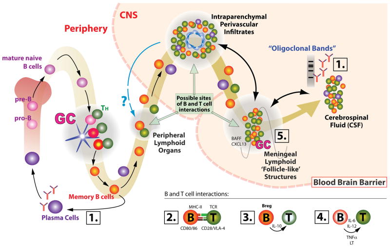
Over the past two decades B cells have increasingly moved into the spotlight in multiple sclerosis (MS) research. This interest was fuelled by growing understanding and acceptance of pathological involvement of B cells and antibodies in MS. Data derived from animal models of MS, human histopathological studies, and analyses of B cells in the peripheral blood and cerebrospinal fluid (CSF) have permitted the integration of B cells in our overall picture of MS immunopathogenesis. The as yet strongest direct evidence for a central role of B cells in MS autoimmunity was the demonstration that peripheral B cell depletion leads to a rapid decline of disease-activity in MS. While lending formidable impact to peripheral blood B cells as mediators of disease activity, the effects of anti-CD20 treatment also seemingly challenged the paradigm of a role of antibodies in targeted central nervous system (CNS) myelin destruction. This review shall attempt to provide an overview of our current understanding of B cell and antibody mediated mechanisms relevant to MS. We will include findings from, both, human studies, and animal models to highlight the complexity of B cell function as it pertains to MS. B cells appear to be effective drivers of inflammatory activity in MS by way of a diverse toolset of cellular functions. These functions appear to be closely linked to B cells that can be found in the periphery. However, by serving as the source of antibodies, B cells offer a direct humoral response that may target the CNS and lead to tissue specific destruction. Therefore, B cells participate in MS pathogenesis on both sides of the blood-brain barrier.
Copyright © 2011 Elsevier Ltd. All rights reserved.
Figures
References
-
- Maloney DG, Liles TM, Czerwinski DK, Waldichuk C, Rosenberg J, Grillo-Lopez A, Levy R. Phase I clinical trial using escalating single-dose infusion of chimeric anti-CD20 monoclonal antibody (IDEC-C2B8) in patients with recurrent B-cell lymphoma. Blood. 1994;84:2457–2466. - PubMed
-
- Leandro MJ, Cambridge G, Ehrenstein MR, Edwards JC. Reconstitution of peripheral blood B cells after depletion with rituximab in patients with rheumatoid arthritis. Arthritis Rheum. 2006;54:613–620. - PubMed
-
- Hauser SL, Waubant E, Arnold DL, Vollmer T, Antel J, Fox RJ, Bar-Or A, Panzara M, Sarkar N, Agarwal S, et al. B-cell depletion with rituximab in relapsing-remitting multiple sclerosis. N Engl J Med. 2008;358:676–688. In this randomized, double-blind, 48-week phase II trial evaluating B cell depletion as therapeutic option in MS, the authors found rapid reduction of markers of disease activity. Results regarding MRI-measures (primary outcome) were highly significant; results regarding clinical disease activity (secondary outcome) were also significant despite the short study duration. This is the first and largest study to clearly show this effect. To date, this study, along with smaller studies, provides the most important direct evidence for B cell involvement in MS. - PubMed
-
- del Martin MP, Cravens PD, Winger R, Kieseier BC, Cepok S, Eagar TN, Zamvil SS, Weber MS, Frohman EM, Kleinschmidt-Demasters BK, et al. Depletion of B lymphocytes from cerebral perivascular spaces by rituximab. Arch Neurol. 2009;66:1016–1020. - PubMed
Publication types
MeSH terms
Substances
Grants and funding
LinkOut - more resources
Full Text Sources
Other Literature Sources
Medical
Miscellaneous
