. 2011 Oct 9;13(11):1361-7.
doi: 10.1038/ncb2354.

Microtubules induce self-organization of polarized PAR domains in Caenorhabditis elegans zygotes

Affiliations

Microtubules induce self-organization of polarized PAR domains in Caenorhabditis elegans zygotes

Fumio Motegi et al. Nat Cell Biol. .

Abstract

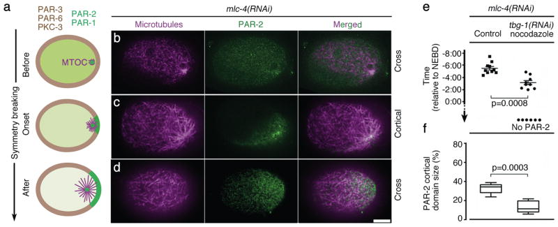
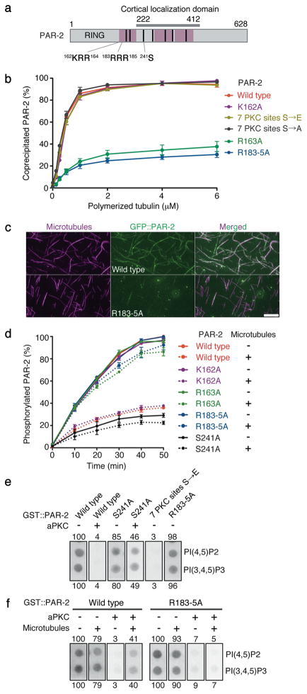
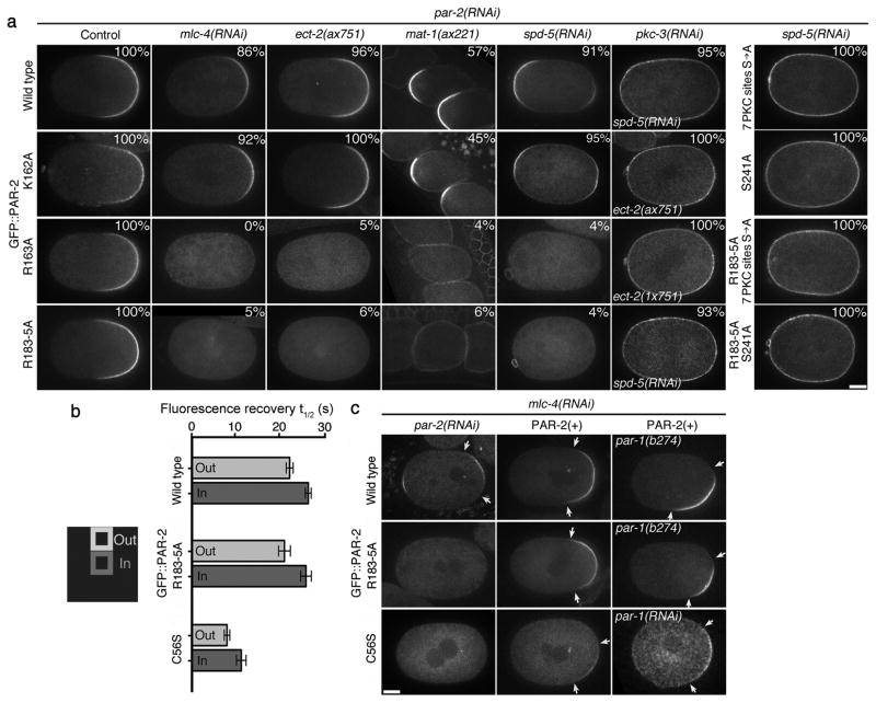
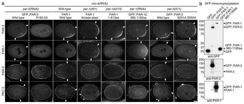
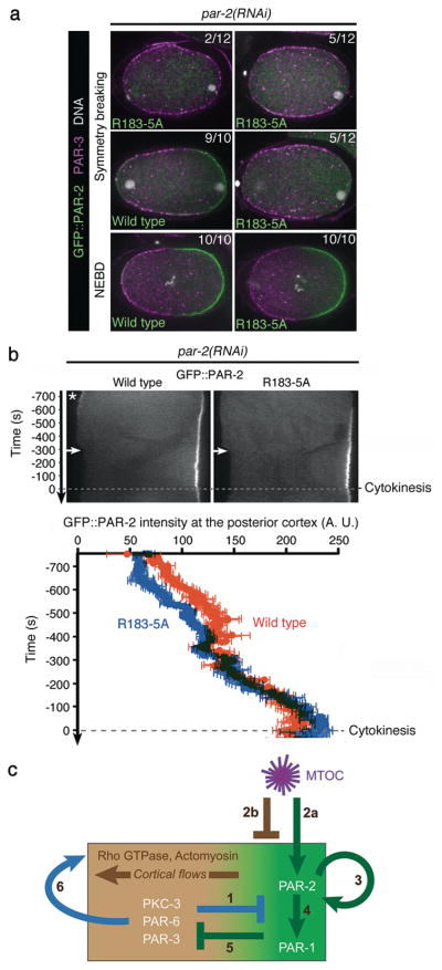
A hallmark of polarized cells is the segregation of the PAR polarity regulators into asymmetric domains at the cell cortex. Antagonistic interactions involving two conserved kinases, atypical protein kinase C (aPKC) and PAR-1, have been implicated in polarity maintenance, but the mechanisms that initiate the formation of asymmetric PAR domains are not understood. Here, we describe one pathway used by the sperm-donated centrosome to polarize the PAR proteins in Caenorhabditis elegans zygotes. Before polarization, cortical aPKC excludes PAR-1 kinase and its binding partner PAR-2 by phosphorylation. During symmetry breaking, microtubules nucleated by the centrosome locally protect PAR-2 from phosphorylation by aPKC, allowing PAR-2 and PAR-1 to access the cortex nearest the centrosome. Cortical PAR-1 phosphorylates PAR-3, causing the PAR-3-aPKC complex to leave the cortex. Our findings illustrate how microtubules, independently of actin dynamics, stimulate the self-organization of PAR proteins by providing local protection against a global barrier imposed by aPKC.


Figures

Figure 1
Figure 1. PAR-2 dynamics at symmetry breaking
(a) Embryo schematics showing the distribution of PAR-1 and PAR-2 (green), anterior PARs (brown), and MTOC/microtubules (magenta). Zygotes are oriented with posterior to the right in this and all figures. (b–d) Confocal images of fixed mlc-4(RNAi) zygotes stained for tubulin (magenta) and PAR-2 (green). Note that b shows a cross-section as in the schematics in a, whereas c and d show superficial cortical sections. Scale bar, 10 μm. (e) Graph showing the timing of GFP::PAR-2 appearance on the posterior cortex in live mlc-4(RNAi) zygotes relative to nuclear envelope breakdown (NEBD). Each dot represents an individual zygote. “no PAR-2” refers to zygotes where PAR-2 never loaded on the cortex. “tbg-1(RNAi) nocodazole” refers to zygotes depleted for gamma-tubulin and treated with nocodazole. Error bars represent standard deviation from 10 control zygotes and 9 tbg-1(RNAi) nocodazole zygotes with a cortical GFP::PAR-2 domain. (f) Graphs showing size of GFP::PAR-2 domain scored at NEBD. Error bars represent standard deviation in zygotes with a cortical GFP::PAR-2 domain as in e. See Supplementary Fig. S1d for images of zygotes used to compile data in e and f.
Figure 2
Figure 2. Microtubule binding protects PAR-2 from aPKC phosphorylation and allows PAR-2 to interact with phospholipids in the presence of aPKC
(a) PAR-2 schematic. Pink boxes are regions that contribute to microtubule binding in vitro (see Supplementary Fig. S3a). Cortical localization domain is the region sufficient for localization to the posterior cortex in the presence of endogenous PAR-2 (reference and F. Motegi, unpublished). Black bars indicate seven potential PKC-3 phosphorylation sites. S241 is required for maximal phosphorylation in vitro by aPKC (Fig. 2d) and for cortical exclusion in vivo (Fig. 3a). 162KRR164 is the basic cluster mutated in the single substitution mutants K162A and R163A, and 183RRR185 is the basic cluster mutated in the triple substitution mutant R183-5A. (b) Graph depicting the percent recombinant PAR-2 that co-sedimented with microtubules. Error bars represent standard deviation in three independent experiments. (c) Photomicrographs of recombinant GFP::PAR-2 mixed with rhodamine-labeled microtubules and spread on slides. GFP::PAR-2(R183-5A) does not decorate microtubules as efficiently as wild-type GFP::PAR-2. Scale bar, 5 μm. (d) Graph depicting the percent phosphorylated PAR-2 with respect to time since start of incubation with aPKC kinase in the presence (dotted lines) or absence (solid lines) of microtubules. PAR-2 phosphorylation was monitored by 32P-ATP incorporation. Error bars represent standard deviation in three independent experiments. (e) Phosphorylation by aPKC inhibits PAR-2 from binding to phospholipids. GST::PAR-2 fusions pre-treated with or without aPKC were incubated with lipid strips and detected using an anti-GST antibody. 50 pmol PI(4,5)P2 and PI(3,4,5)P3 spots are shown (see Supplementary Fig. S7b for the full dilution series). Numbers represent % binding normalized to wild type (100%). S241 is one of 7 predicted aPKC sites. 7PKCsitesS→E is a phosphomimic mutant for all seven sites. (f) Binding to microtubules is sufficient to protect PAR-2 from aPKC and retain binding to phospholipids. Same as in e, but GST::PAR-2 fusions were incubated with microtubules before incubation with aPKC. See Supplementary Fig. S7c for the full dilution series.
Figure 3
Figure 3. Microtubule binding is required for PAR-2 to localize to the cortex in the absence of cortical flows
(a) Live zygotes expressing the indicated GFP::PAR-2 fusions: wild type and K162A bind microtubules, R163A and R183-5A do not. % indicate zygotes with cortical PAR-2, numbers are presented in Supplementary Table 1. ECT-2 is the GEF for the small GTPase RHO-1. ect-2(ax751) lack MTOC-induced cortical flows, but develop PAR-2-dependent cortical flows during mitosis. MAT-1 is a subunit of the anaphase-promoting complex. mat-1(ax227) zygotes arrest in meiosis and become transiently polarized without cortical flows under the influence of the acentriolar meiotic spindle. SPD-5 is a MTOC component required for PCM assembly. spd-5(RNAi) zygotes localize GFP::PAR-2 to both the anterior and posterior cortex under the influence of the meiotic spindle remnant (anterior) and the slow maturing MTOC (posterior). RNAi depletion of PKC-3 or mutations in the PKC phosphorylation sites (either 7 PKC sites S→A or S241A) causes all fusions to localize uniformly to the cortex. Scale bar, 10 μm. (b) Fluorescence Recovery After Photobleaching was performed on the cortex of pkc-3(RNAi) zygotes expressing the indicated GFP::PAR-2 fusions. Graph shows the average recovery half time (t½) from five separate zygotes. Error bars represent standard deviation. Fluorescence recovery was faster at the boundary (Out) than at the center (In) of the bleached area, suggesting that at least some of the recovery is due to lateral diffusion of cortical GFP::PAR-2 as shown in reference. See supplementary Fig. S5b for representative recovery curves. (c) Cortical PAR-2 stimulates its own recruitment to the cortex. Live zygotes expressing the indicated GFP::PAR-2 fusions. Arrows point to the boundaries of the cortical GFP::PAR-2 domain. Scale bar, 10 μm. In mlc-4(RNAi);par-2(RNAi) zygotes, wild-type PAR-2 localizes to the posterior cortex, but the microtubule-binding mutant R183-5A and the RING mutant C56S do not. Endogenous PAR-2 [PAR-2(+)] rescues the localization of both mutants. Rescue is also observed in par-1 zygotes, where PAR-3 and PKC-3 are never excluded from the posterior cortex (see Fig. 4a).
Figure 4
Figure 4. PAR-2 recruits PAR-1 to the cortex, leading to exclusion of anterior PARs
(a) mlc-4(RNAi) zygotes with indicated mutations in PAR proteins stained for PAR-2, PAR-1, PAR-3 and PKC-3. par-1(it51) contains a mutation (R409K) that inhibits kinase activity, and par-1(b274) contains a premature stop (Q814Stop) that eliminates the PAR-1 cortical localization domain. GFP::PAR-3(S251A S950A) contains mutations in the conserved PAR-1 phosphorylation sites and rescues par-3(it71) zygotes competent for cortical flows. GFP::PAR-2 fusions were co-stained with PAR-1. PAR-2 and PKC-3 or PAR-1 and PAR-3 were co-stained in the other zygotes. Arrows indicate the boundary of the PAR domains. Scale bar, 10 μm. (b) Immunoprecipitation experiment showing that PAR-2 and PAR-1 interact in embryo extracts. Extracts from embryos expressing the indicated GFP fusions were immunoprecipitated with anti-GFP-beads and the immunoprecipitates were blotted with the indicated antibodies.
Figure 5
Figure 5. Microtubule binding by PAR-2 is required for efficient polarity initiation in wild-type embryos
(a) Fluorescent micrographs of fixed zygotes expressing GFP::PAR-2 and depleted for endogenous PAR-2 by RNAi. Zygotes are stained for GFP::PAR-2 (green), PAR-3 (magenta), and DNA (white) and shown at symmetry breaking (first two rows) or at NEBD (last row). Scale bar, 10 μm. (b) Kymographs from time-lapse movies of live zygotes expressing GFP::PAR-2 fusions and depleted for endogenous PAR-2 by RNAi. Times are with respect to the onset of cytokinesis. Wild-type GFP::PAR-2 appears on the posterior cortex earlier than the microtubule-binding mutant GFP::PAR-2(R183-5A). Wild-type GFP::PAR-2 also accumulates transiently (asterisk) on the anterior cortex (due to the transient influence of the meiotic spindle remnant; 5 of 5 zygotes). GFP::PAR-2(R183-5A) does not show this localization (0 of 5), consistent with polarization by the meiotic spindle depending primarily on microtubules. Graph shows fluorescence intensity at posterior most cortex averaged from five zygotes. Accumulation of GFP::PAR-2(R183-5A) is delayed compared to wild-type GFP::PAR-2 (29.0 +/− 11.2 seconds, p=0.03) but catches up by NEBD. Error bars represent standard deviation from five separate zygotes. (c) Model for polarization of the C. elegans zygote 1) PKC-3 phosphorylates PAR-2 and PAR-1, keeping them off the cortex. 2) MTOC breaks symmetry via two parallel mechanisms: 2a; Microtubules at the MTOC protect PAR-2 from phosphorylation by PKC-3, allowing a few molecules of PAR-2 to load on cortex close to MTOC. 2b; MTOC induces cortical flows by an unknown mechanism involving local inhibition of actomyosin. Flows displace anterior PARs, allowing PAR-2 to accumulate in their place. 3) Cortical PAR-2 recruits additional PAR-2 molecules to expand the PAR-2 domain. RING finger of PAR-2 stabilizes PAR-2 at the cortex. 4) PAR-2 recruits PAR-1 by binding to the C-terminus of PAR-1. 5) PAR-1 phosphorylates PAR-3 preventing its association with the cortex. 6) Anterior PARs stimulate their own displacement by recruiting myosin to the cortex and up-regulating cortical flows, . Not shown in this figure is LGL, a non-essential player in this process, which like PAR-1 localizes to the posterior cortex and antagonizes the cortical association of anterior PARs, .

Comment in

References

    1. Goldstein B, Macara I. The PAR proteins: fundamental players in animal cell polarization. Dev Cell. 2007;13:609–622. - PMC - PubMed
    1. St Johnston D, Ahringer J. Cell polarity in eggs and epithelia: parallels and diversity. Cell. 2010;141:757–774. - PubMed
    1. Munro E, Nance J, Priess JR. Cortical flows powered by asymmetrical contraction transport PAR proteins to establish and maintain anterior-posterior polarity in the early C. elegans embryo. Dev Cell. 2004;7:413–424. - PubMed
    1. Zonies S, Motegi F, Hao Y, Seydoux G. Symmetry breaking and polarization of the C. elegans zygote by the polarity protein PAR-2. Development. 2010;137:1669–1677. - PMC - PubMed
    1. Cuenca AA, Schetter A, Aceto D, Kemphues K, Seydoux G. Polarization of the C. elegans zygote proceeds via distinct establishment and maintenance phases. Development. 2003;130:1255–1265. - PMC - PubMed

Publication types

MeSH terms