

doi: 10.1038/nn.2934.
Sleep and waking modulate spine turnover in the adolescent mouse cortex
Affiliations
- PMID: 21983682
- PMCID: PMC3203346
- DOI: 10.1038/nn.2934
Item in Clipboard
Sleep and waking modulate spine turnover in the adolescent mouse cortex
Nat Neurosci.
.
Abstract
Cortical development involves synaptic formation and elimination. Although synaptogenesis predominates in the early stages and pruning in the later stages, the two processes are thought to happen concurrently. In adults, synaptic strength is modulated by behavioral state, and we asked whether synaptic remodeling may be affected by sleep and waking states. Using two-photon microscopy in adolescent mice, we found that waking results in a net increase in cortical spines, whereas sleep is associated with net spine loss.
Figures
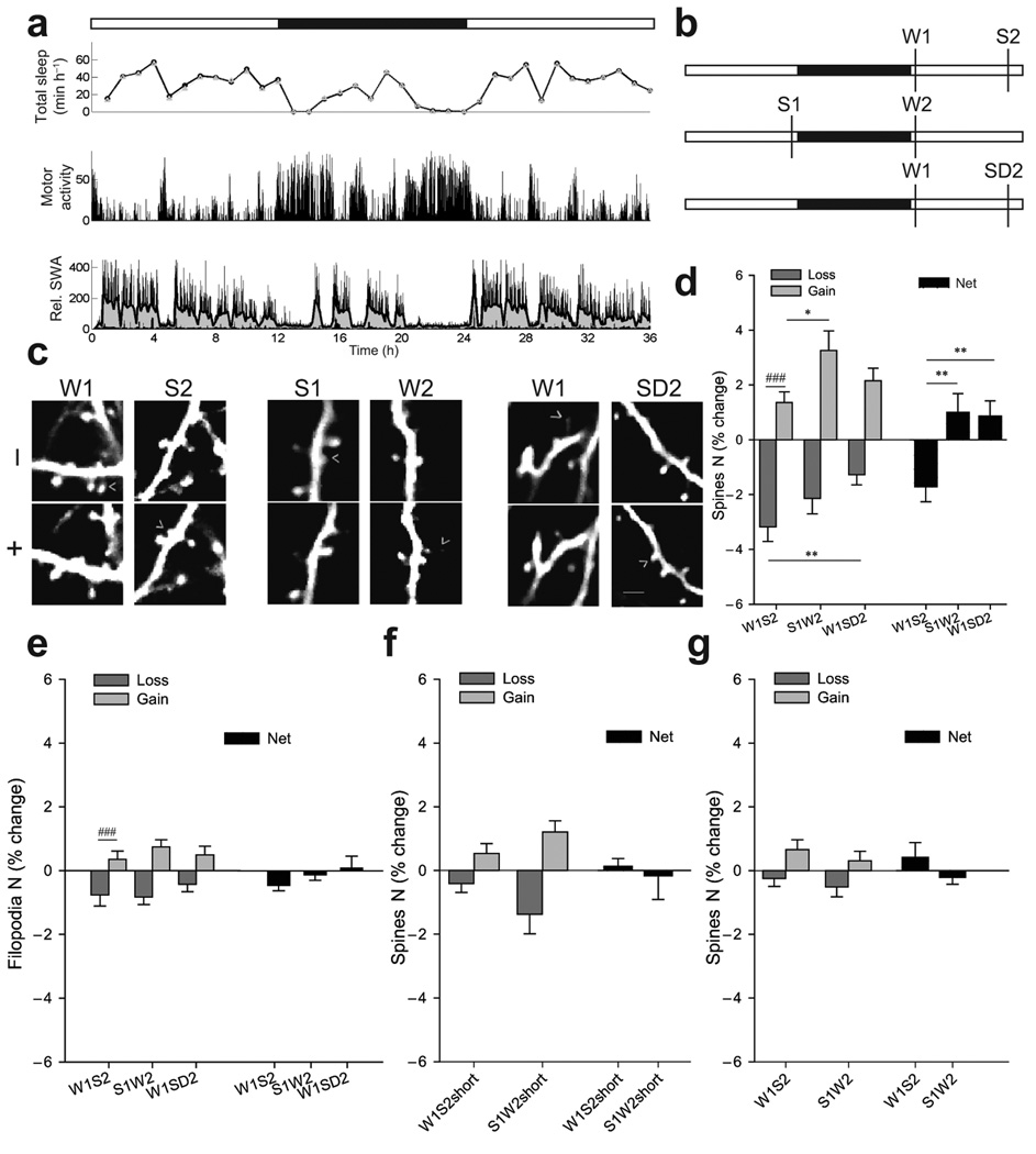
(a) Sleep/wake pattern in one P30 mouse. White and black bars indicate light and dark phase, respectively. Upper panel, total sleep time based on EEG recordings (circles) or video recording of motor activity (triangles). Middle panel, motor activity detected by video recording. Lower panel, slow wave activity (SWA, 0.5–4Hz EEG power in NREM sleep, % of 24hrs), an index of NREM sleep intensity. Gray area shows the moving average of ten 4-sec epochs. In ~P30 mice SWA is homeostatically regulated, being high at the beginning of the light phase and declining during sleep. (b) Experimental groups. (c) Repeated imaging of the same dendrite with examples of spine formation and loss (arrowheads; 1 filopodium is shown in W1). Scale bar, 6µm. (d) Spines formed and lost (% of all spines in first session) and net change (mean±SEM; intergroup, *p<0.05, **p<0.01, Kruskal-Wallis; intragroup, ### p<0.001, Wilcoxon). (e) Changes in filopodia (% of all protrusions in first session; mean±SEM; ### p<0.001, Wilcoxon). (f–g) Spine changes after short sleep/wake and in adults.
References
Publication types
MeSH terms
Substances
Grants and funding
LinkOut - more resources
Full Text Sources
