

Tracheal dimensions in human fetuses: an anatomical, digital and statistical study
- PMID: 21984196
- PMCID: PMC3334485
- DOI: 10.1007/s00276-011-0878-7
Tracheal dimensions in human fetuses: an anatomical, digital and statistical study
Abstract
Purpose: Rapid advances in perinatal medicine have resulted in increased number of various tracheo-bronchial interventions on fetal and neonatal airways. The present study was performed to compile normative data for external dimensions of the trachea at varying gestational age.
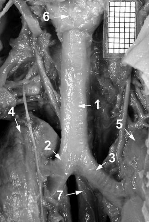
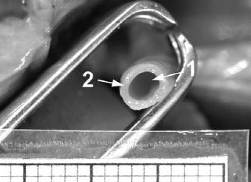
Materials and methods: Using anatomical dissection, digital image analysis (NIS-Elements BR 3.0) and statistical analysis (ANOVA, regression analysis), a range of measurements (prebifurcation and bifurcation lengths, proximal and distal external transverse diameters, proximal external cross-sectional area, and external volume) for the trachea in 73 spontaneously aborted fetuses (39 male, 34 female) aged 14-25 weeks was examined.
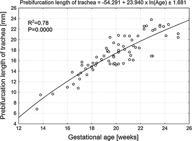
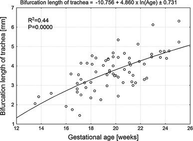
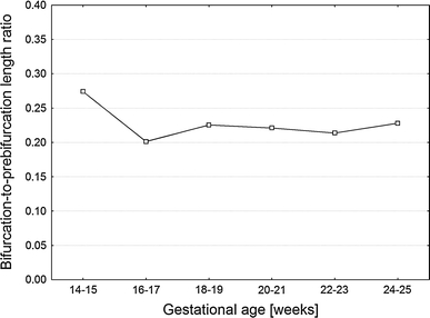
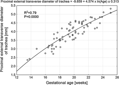
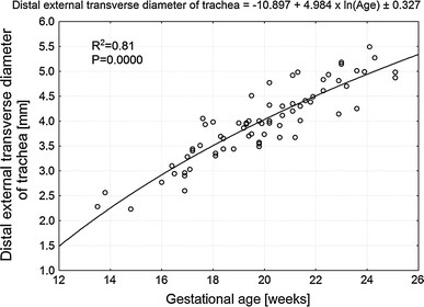
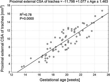
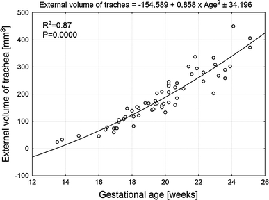
Results: No significant male-female differences were found (P > 0.05). The prebifurcation and bifurcation lengths ranged from 8.14 ± 1.90 to 20.77 ± 0.50 mm and from 2.23 ± 0.25 to 5.77 ± 0.76 mm, according to the functions y = -54.291 + 23.940 × ln (Age) ± 1.681 (R (2) = 0.78) and y = -10.756 + 4.860 × ln (Age) ± 0.731 (R (2) = 0.44), respectively. Their relative growth, expressed as the bifurcation-to-prebifurcation length ratio, was stable from the age of 16 weeks and attained the value 0.22 ± 0.05. The proximal external transverse diameter of the trachea was greater (36 fetuses, 49.3%), smaller (34 fetuses, 46.6%) or similar (3 fetuses, 4.1%), when compared to the distal external transverse diameter. The values for proximal and distal transverse diameters ranged from 2.39 ± 0.04 to 5.20 ± 0.17 mm and from 2.42 ± 0.20 to 4.93 ± 0.08 mm, expressed by the functions: y = -9.659 + 4.574 × ln (Age) ± 0.313 (R (2) = 0.79) and y = -10.897 + 4.984 × ln (Age) ± 0.327 (R (2) = 0.81). The values of proximal external cross-sectional area ranged from 3.38 ± 0.12 to 15.98 ± 1.04 mm(2), according to the linear function y = -11.798 + 1.077 × Age ± 1.463 (R (2) = 0.78). The values of external volume of the trachea ranged from 34.3 ± 11.6 to 370.6 ± 94.1 mm(3) and generated the quadratic function y = -154.589 + 0.858 × Age(2) ± 34.196 (R (2) = 0.87).
Conclusions: The tracheal parameters do not show male-female differences. The developmental dynamics of prebifurcation and bifurcation lengths and proximal and distal external transverse diameters of the trachea follow linear functions dependent on the natural logarithm of fetal age, its external cross-sectional area-according to a linear function, and its external volume-according to a quadratic function.
© The Author(s) 2011. This article is published with open access at Springerlink.com
Figures
Similar articles
-
New anatomical data on the growing C4 vertebra and its three ossification centers in human fetuses.Surg Radiol Anat. 2013 Apr;35(3):191-203. doi: 10.1007/s00276-012-1022-z. Epub 2012 Sep 18. Surg Radiol Anat. 2013. PMID: 22986651 Free PMC article.
-
New quantitative patterns of the growing trachea in human fetuses.Med Sci Monit. 2012 Jun;18(6):PH63-70. doi: 10.12659/msm.882890. Med Sci Monit. 2012. PMID: 22648261 Free PMC article.
-
Quantitative anatomy of the growing abdominal aorta in human fetuses: an anatomical, digital and statistical study.Med Sci Monit. 2012 Oct;18(10):BR419-26. doi: 10.12659/msm.883483. Med Sci Monit. 2012. PMID: 23018350 Free PMC article.
-
Morphometric study of the T6 vertebra and its three ossification centers in the human fetus.Surg Radiol Anat. 2013 Dec;35(10):901-16. doi: 10.1007/s00276-013-1107-3. Epub 2013 Mar 30. Surg Radiol Anat. 2013. PMID: 23543237 Free PMC article.
-
The normal growth of the tracheal wall in human foetuses.Arch Med Sci. 2013 Oct 31;9(5):922-9. doi: 10.5114/aoms.2012.31411. Epub 2012 Oct 30. Arch Med Sci. 2013. PMID: 24273580 Free PMC article.
Cited by
-
Cross-sectional study of the neural ossification centers of vertebrae C1-S5 in the human fetus.Surg Radiol Anat. 2013 Oct;35(8):701-11. doi: 10.1007/s00276-013-1093-5. Epub 2013 Feb 28. Surg Radiol Anat. 2013. PMID: 23455365 Free PMC article.
-
Morphometric study of the diaphragmatic surface of the liver in the human fetus.PLoS One. 2020 Jan 24;15(1):e0227872. doi: 10.1371/journal.pone.0227872. eCollection 2020. PLoS One. 2020. PMID: 31978157 Free PMC article.
-
New anatomical data on the growing C4 vertebra and its three ossification centers in human fetuses.Surg Radiol Anat. 2013 Apr;35(3):191-203. doi: 10.1007/s00276-012-1022-z. Epub 2012 Sep 18. Surg Radiol Anat. 2013. PMID: 22986651 Free PMC article.
-
Tracheo-bronchial angles in the human fetus -- an anatomical, digital, and statistical study.Med Sci Monit Basic Res. 2013 Jul 16;19:194-200. doi: 10.12659/MSMBR.889085. Med Sci Monit Basic Res. 2013. PMID: 23857411 Free PMC article.
-
Novel patterns for the growing main bronchi in the human fetus: an anatomical, digital and statistical study.Surg Radiol Anat. 2014 Jan;36(1):55-65. doi: 10.1007/s00276-013-1145-x. Epub 2013 Jun 19. Surg Radiol Anat. 2014. PMID: 23778946 Free PMC article.
References
MeSH terms
LinkOut - more resources
Full Text Sources
