. 2011 Oct 9;18(11):1204-10.
doi: 10.1038/nsmb.2139.

Crystal structures of the extracellular domain of LRP6 and its complex with DKK1

Affiliations

Crystal structures of the extracellular domain of LRP6 and its complex with DKK1

Zhihong Cheng et al. Nat Struct Mol Biol. .

Abstract

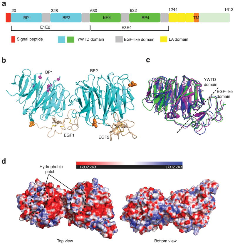
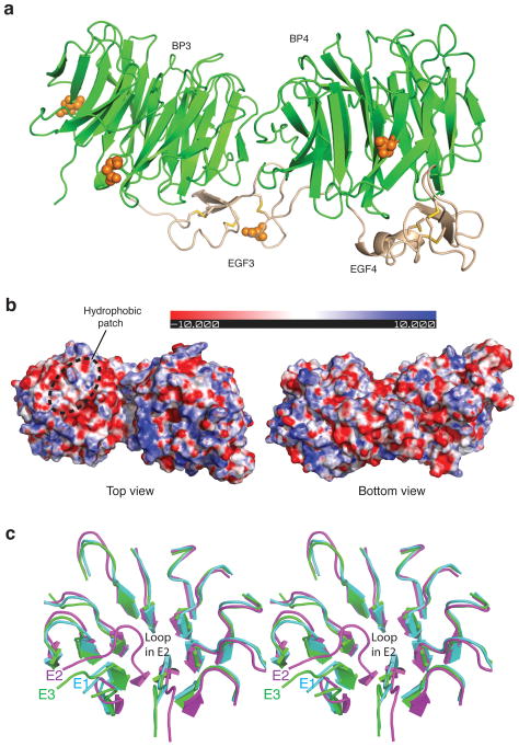
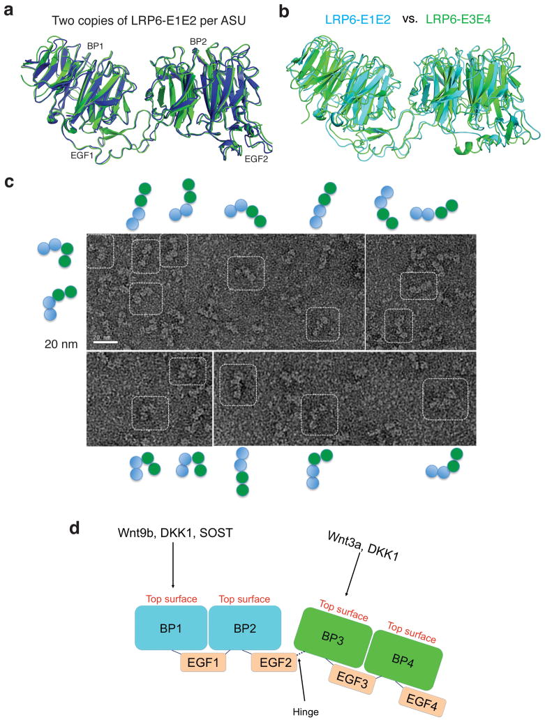
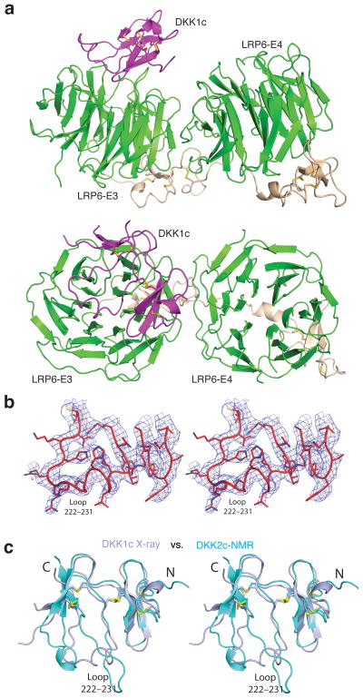
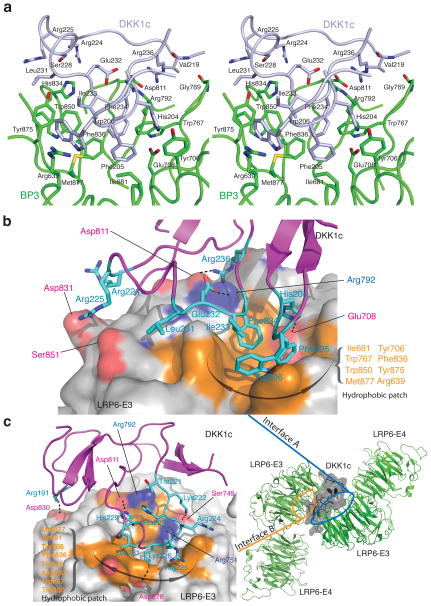
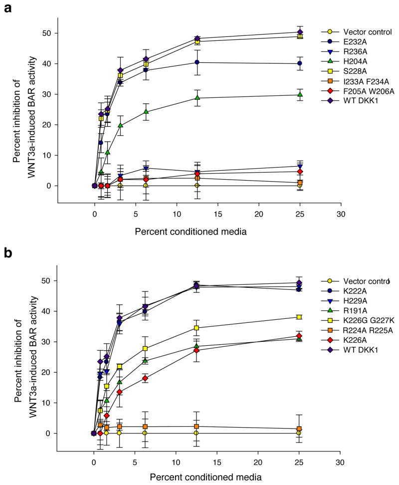
Low-density-lipoprotein (LDL) receptor-related proteins 5 and 6 (LRP5/6) are Wnt co-receptors essential for Wnt/β-catenin signaling. Dickkopf 1 (DKK1) inhibits Wnt signaling by interacting with the extracellular domains of LRP5/6 and is a drug target for multiple diseases. Here we present the crystal structures of a human LRP6-E3E4-DKK1 complex and the first and second halves of human LRP6's four propeller-epidermal growth factor (EGF) pairs (LRP6-E1E2 and LRP6-E3E4). Combined with EM analysis, these data demonstrate that LRP6-E1E2 and LRP6-E3E4 form two rigid structural blocks, with a short intervening hinge that restrains their relative orientation. The C-terminal domain of DKK1 (DKK1c) interacts with the top surface of the LRP6-E3 YWTD propeller and given their structural similarity, probably also that of the LRP6-E1 propeller, through conserved hydrophobic patches buttressed by a network of salt bridges and hydrogen bonds. Our work provides key insights for understanding LRP5/6 structure and the interaction of LRP5/6 with DKK, as well as for drug discovery.


Conflict of interest statement

Competing Financial interests

R.T.M. is a cofounder of, and consultant with, FATE Therapeutics. The other authors declare no competing financial interests.

Figures

Figure 1
Figure 1
Crystal structure of LRP6-E1E2. (a) Schematic representation of the domain organization of human LRP6. “BP” stands for YWTD β-propeller domain. First and second YWTD domains are colored in cyan. Third and fourth YWTD domains are shown in green. EGF-like domains, LDLR type A repeats, and transmembrane region are shown in gray, yellow, and orange, respectively. (b) Overall structure of LRP6-E1E2 fragment. YWTD domains are shown in cyan, EGF-like domain in wheat. Observed N-glycosylation sites/moiety (in orange spheres), and four sites corresponding to LRP5 mutants associated with high bone density syndrome (in magenta balls) are shown. (c) Superposition of three YWTD–EGF pairs: LRP6-E1, LRP6-E2 and the LDLR YWTD–EGF3 pair. LRP6-E1 and LRP6-E2 are shown in magenta and green, respectively, while LDLR is in blue. (d) Electrostatic surfaces of LRP6-E1E2 (top and bottom views).
Figure 2
Figure 2
Crystal structure of LRP6-E3E4. (a) Overall structure and observed N-glycosylation sites. YWTD domains are shown in green and EGF-like domain in wheat. N-glycosylation sites are shown in orange spheres. (b) Electrostatic surfaces of LRP6-E3E4 (top and bottom views). (c) Comparison of the top surfaces of LRP6-E3 vs E2, and E3 vs E1. Structures of LRP6-E3 and E1, E2 are superimposed. It is clear that LRP6-E1, but not LRP6-E2, has a very similar top surface with LRP6-E3. The difference in the E2-propeller top surface is mainly due to the “bulge out” of the loop between blades 5 and 6, which is observed in both copies of LRP6-E2.
Figure 3
Figure 3
Rigidity of the two LRP6-ECD structural blocks and hinge between these two blocks. (a) Superposition of two LRP6-E1E2 copies in the asymmetric unit of the LRP6-E1E2 crystal lattice. (b) Superposition of LRP6-E3E4 and LRP6-E1E2. LRP6-E3E4 and E1E2 are shown in green and cyan, respectively. (c) EM analysis of human LRP6-E1E4. (d) A model for the two structural blocks of the LRP6 extracellular domain and the hinge between these two blocks.
Figure 4
Figure 4
Crystal structure of the LRP6-E3E4/DKK1c complex. (a) An overall view of the complex in two orientations roughly related by a horizontal 90° rotation. Only the interface A is shown here (see text). DKK1c is in magenta. (b) Stereo view of a representative portion of the Fo-Fc electron density map (contoured at 2.0 σ), showing the DKK1c loop 222–231 region. The map was calculated using phases from LRP6-E3E4 only. (c) Comparison of DKK1c and DKK2c structures. Crystal structure of DKK1 in the LRP6-E3E4/DKK1 complex (in light blue) and the NMR structure of DKK2c (in cyan) are superimposed.
Figure 5
Figure 5
Specific interactions in the two LRP6-E3/DKK1c interfaces. (a) Stereo view of LRP6-E3E4/DKK1 interface A. DKK1c is shown in light blue. Residues involved in the interactions are labeled and shown in green (LRP6-E3E4) and light blue (DKK1c) stick models. (b) A surface representation of the LRP6–DKK1c interaction (interface A only). LRP6-E3 is shown in surface illustration. LRP6 residues involved in the interface are shown on the solid surface, whereas DKK1 residues are shown in stick models. Hydrophobic LRP6 and DKK1c residues directly involved in hydrophobic interactions are also labeled in orange and cyan, respectively. (c) The second LRP6–DKK1 interface (interface B) observed in the crystal lattice. (Right) Overall structure of LRP6-E3E4/DKK1c complex in the crystal lattice. Two LRP6-E3E4 are arranged roughly in an anti-parallel orientation. DKK1c is shown in transparent surface illustration. (Left) Detailed interactions on the interface B. LRP6-E3 is shown in surface illustration and its interface residues are shown as in (b). The physiological relevance of the interface B remains to be tested.
Figure 6
Figure 6
DKK1 residues in DKK1–LRP5/6 interface are important for its inhibitory effects on Wnt/β-catenin signaling. Conditioned media was generated for each of the indicated DKK1 constructs and each was tested for its ability to inhibit Wnt3A mediatedβ-catenin signaling in RKO cells as measured by a β-catenin activated reporter (BAR). (a) mutations on interface A; (b) mutations on interface B. Assays were performed in triplicate and error bars represent standard deviation. Reporter data were normalized to expression of the corresponding DKK1 WT or mutant protein as determined by desitometric analysis of a western blot (Supplementary Fig. 6).

Comment in

Similar articles

Cited by

References

    1. Reya T, Clevers H. Wnt signalling in stem cells and cancer. Nature. 2005;434:843–50. - PubMed
    1. Moon RT, Bowerman B, Boutros M, Perrimon N. The promise and perils of Wnt signaling through beta-catenin. Science. 2002;296:1644–6. - PubMed
    1. Nelson WJ, Nusse R. Convergence of Wnt, beta-catenin, and cadherin pathways. Science. 2004;303:1483–7. - PMC - PubMed
    1. Petersen CP, Reddien PW. Wnt signaling and the polarity of the primary body axis. Cell. 2009;139:1056–68. - PubMed
    1. MacDonald BT, Tamai K, He X. Wnt/beta-catenin signaling: components, mechanisms, and diseases. Dev Cell. 2009;17:9–26. - PMC - PubMed

Publication types

MeSH terms

Substances