. 2011 Oct 9;18(11):1196-203.
doi: 10.1038/nsmb.2128.

Structure and nucleosome interaction of the yeast NuA4 and Piccolo-NuA4 histone acetyltransferase complexes

Affiliations

Structure and nucleosome interaction of the yeast NuA4 and Piccolo-NuA4 histone acetyltransferase complexes

Johnathan R Chittuluru et al. Nat Struct Mol Biol. .

Abstract

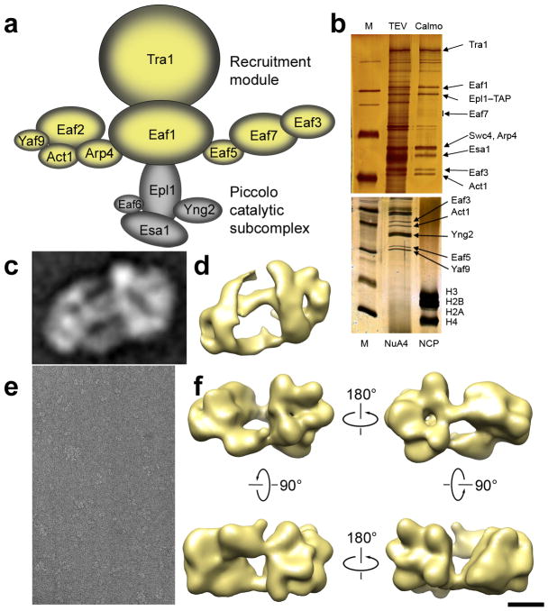
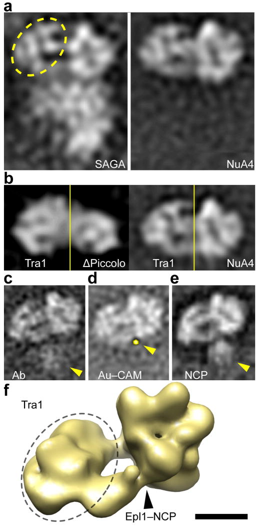
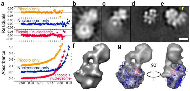
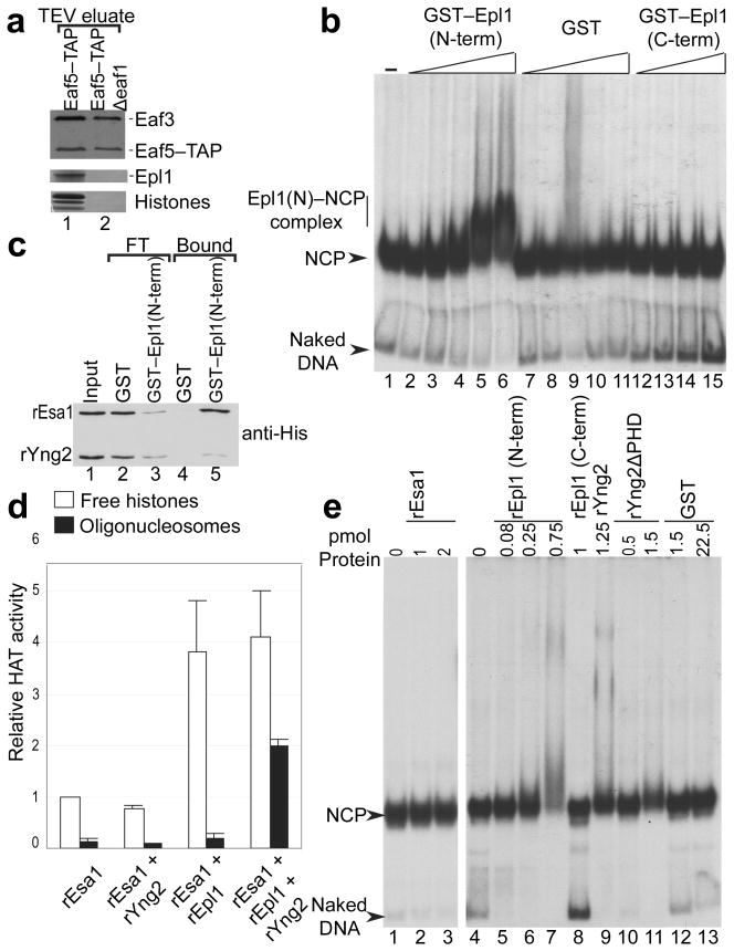
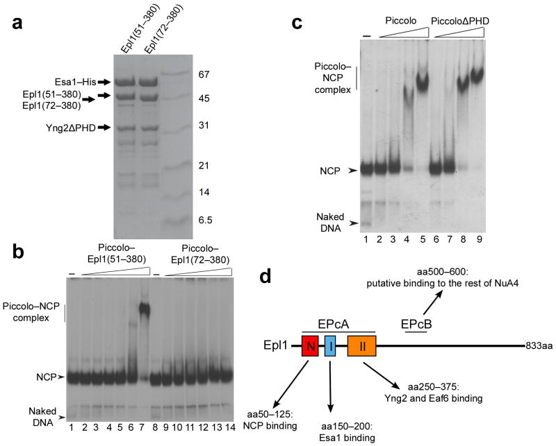
We have used EM and biochemistry to characterize the structure of NuA4, an essential yeast histone acetyltransferase (HAT) complex conserved throughout eukaryotes, and we have determined the interaction of NuA4 with the nucleosome core particle (NCP). The ATM-related Tra1 subunit, which is shared with the SAGA coactivator complex, forms a large domain joined to a second region that accommodates the catalytic subcomplex Piccolo and other NuA4 subunits. EM analysis of a NuA4-NCP complex shows the NCP bound at the periphery of NuA4. EM characterization of Piccolo and Piccolo-NCP provided further information about subunit organization and confirmed that histone acetylation requires minimal contact with the NCP. A small conserved region at the N terminus of Piccolo subunit enhancer of Polycomb-like 1 (Epl1) is essential for NCP interaction, whereas the subunit yeast homolog of mammalian Ing1 2 (Yng2) apparently positions Piccolo for efficient acetylation of histone H4 or histone H2A tails. Taken together, these results provide an understanding of the NuA4 subunit organization and the NuA4-NCP interactions.


Figures

Figure 1
Figure 1. NuA4 subunit organization and EM characterization
(a) Model for the organization of NuA4 subunits into functional modules arranged around the Eaf1 scaffold protein (based on). Subunits involved in recruitment of NuA4 to specific loci are colored yellow, and subunits comprising the Piccolo catalytic subcomplex are colored gray. (b) SDS–PAGE analysis of NuA4 after TAP purification. The lower gel shows that no histones copurify with NuA4. Subunit Eaf6 (∼16 kDa) is not visible since it stains very poorly with silver. (c) A 2D class average of NuA4 obtained by reference-free alignment of EM images of particles preserved in stain. (d) A view (corresponding to the projection shown in panel b) of a 3D reconstruction of NuA4 obtained using the RCT method. The 2D and 3D maps both show a structure comprising two large domains joined by thin connections. (e) Representative electron micrograph of NuA4 particles preserved in amorphous ice. (f) Different views of a cryo-EM map of NuA4 calculated from ∼35,000 individual-particle cryo images. The view in the top left corresponds to the orientation preferentially adopted by particles preserved in stain. Scale bars represent 5 nm (c, d, and f) and 100 nm (e).
Figure 2
Figure 2. NuA4 subunit organization, interaction with the nucleosome, and structural homology with SAGA
(a) 2D class averages obtained by reference-free alignment of SAGA (left) and NuA4 (right) stained particle images. The upper portion of the SAGA structure, whose Tra1 portion is highlighted by a dashed ellipse, is remarkably similar to the NuA4 map. The orientation of NuA4 shown here to match the SAGA map corresponds to a ∼20° rotation in the plane of the page from the NuA4 averages presented elsewhere. (b) Projection structure of a ΔPiccolo NuA4 mutant (left) next to a projection of wild-type NuA4 (right). The Tra1 portions of the ΔPiccolo and wild-type NuA4 (to the left of the yellow line) are very similar. In contrast, the non-Tra1 portion (to the right of the yellow line) of the ΔPiccolo map is much smaller, reflecting the loss of Piccolo density. (c) Immunolabeling of the Epl1 subunit with anti-CBP Ab results in disordered antibody density highlighted by the arrowhead. (d) Labeling of the same CBP tag on Epl1 using a calmodulin-derivatized gold cluster results in gold cluster density (yellow arrowhead and false-color overlay highlighting pixels with intensities above 3σ). (e) Incubation of NuA4 with nucleosomes results in localized density (highlighted by arrowhead) at the periphery of NuA4 in a position adjacent to the Epl1 subunit. (f) The portion of the NuA4 structure corresponding to Tra1 (dashed ellipse) and the location of Epl1 and the NCP binding site (black arrowhead) are highlighted in the 3D cryo-EM map. Scale bars represent 10 nm (a, b), 13.5 nm (ce), and 5 nm (f).
Figure 3
Figure 3. Characterization of the Piccolo–NCP complex by AUC and EM
(a). AUC data for Piccolo, NCP, and Piccolo–NCP are shown as orange diamonds, blue triangles, and red circles, respectively, while fitted curves are shown as solid lines. The residuals above document the small difference between the experimental data and the fitted curve used to determine the molecular weights (see Table 1). (b) Class average obtained after reference-free alignment of images of individual Piccolo–NCP particles. (c) Piccolo class average obtained after masking out NCP density in Piccolo–NCP images (d) Piccolo projection map obtained from images of Piccolo alone. (e) Piccolo projection map obtained from images of a different Piccolo deletion variant including additional Epl1 and Yng2 domains. (f) 3D map of the Piccolo–NCP complex obtained by the RCT method. (g) Docking of the X-ray structure of the nucleosome into the Piccolo–NCP 3D map. The H4HFD is highlighted in red. Scale bars represent 5 nm (b) through (e) and 3.5 nm (f, g).
Figure 4
Figure 4. Epl1 N-terminus drives binding to nucleosomes within NuA4, but Yng2 is required for acetylation
(a) Physical association of histones with NuA4 requires the Piccolo subcomplex in vivo. (b) Epl1 N-terminus has strong binding affinity for mononucleosomes. A stable complex is detected between the Epl1 N-terminus and the NCP (lanes 5 and 6). (c) GST pull downs using near stoichiometric ratios of the indicated recombinant proteins were carried out and assayed by western blotting. As shown using anti-His antibody for western blots, both rEsa1 and rYng2 are pulled down using GST–Epl1 (N-term). Note that while rEsa1 and rYng2 are pulled down simultaneously using GST–Epl1 (N-term), rEsa1 is more efficiently brought with Epl1. (d) Relative HAT activity of rEsa1 alone or in combination with other recombinant proteins present in Piccolo. HAT assays were done on free (open bars) or nucleosomal (black bars) histones. Error bars indicate s.d. (e) NCP mobility shift assay showing that the N-termini of rEpl1 and rYng2 are able to interact with nucleosomes while rEsa1 and rEpl1 (C-term) cannot (compare lanes 7 and 9 with 2, 3, and 8).
Figure 5
Figure 5. Epl1 residues 51–72 are essential for Piccolo–NCP interaction
(a) SDS–PAGE analysis of purified Piccolo constructs comprising full-length Esa1, Yng2ΔPHD, and either Epl1(51–380) or Epl1(72–380). (b) Mobility shift assay indicating that Epl1 residues 51–71 are required for the binding interaction between Piccolo and mononucleosomes. (c) Mobility shift assay indicating that the Yng2 PHD domain is dispensable for binding of mononucleosomes by the recombinant Piccolo complex. (d) Diagram illustrating the residues required for known Epl1 binding interactions elucidated by this and previous or parallel studies,. The conserved domain EPcA (residues 50–380) can be subdivided in three conserved amino acids clusters with subdomains I and II being responsible for Piccolo assembly by bridging Esa1 together with Yng2 and the subdomain N being the critical NCP binding surface. Amino acid, aa.

Similar articles

Cited by

References

    1. Allard S, et al. NuA4, an essential transcription adaptor/histone H4 acetyltransferase complex containing Esa1p and the ATM-related cofactor Tra1p. EMBO J. 1999;18:5108–5119. - PMC - PubMed
    1. Clarke AS, Lowell JE, Jacobson SJ, Pillus L. Esa1p is an essential histone acetyltransferase required for cell cycle progression. Mol Cell Biol. 1999;19:2515–2526. - PMC - PubMed
    1. Selleck W, Fortin I, Sermwittayawong D, Cote J, Tan S. The Saccharomyces cerevisiae Piccolo NuA4 histone acetyltransferase complex requires the Enhancer of Polycomb A domain and chromodomain to acetylate nucleosomes. Mol Cell Biol. 2005;25:5535–5542. - PMC - PubMed
    1. Boudreault AA, et al. Yeast enhancer of polycomb defines global Esa1-dependent acetylation of chromatin. Genes Dev. 2003;17:1415–1428. - PMC - PubMed
    1. Avvakumov N, Cote J. The MYST family of histone acetyltransferases and their intimate links to cancer. Oncogene. 2007;26:5395–5407. - PubMed

Publication types

MeSH terms