

Ruminant rhombencephalitis-associated Listeria monocytogenes alleles linked to a multilocus variable-number tandem-repeat analysis complex
- PMID: 21984240
- PMCID: PMC3233052
- DOI: 10.1128/AEM.06507-11
Ruminant rhombencephalitis-associated Listeria monocytogenes alleles linked to a multilocus variable-number tandem-repeat analysis complex
Abstract
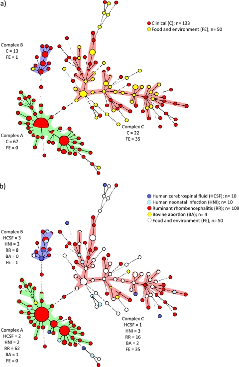
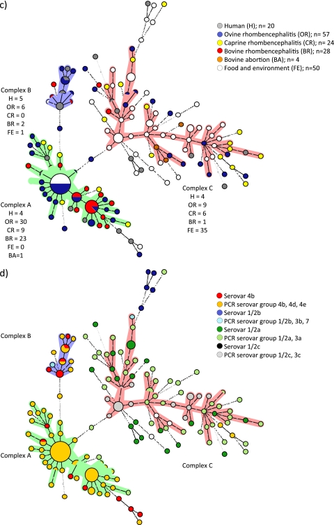
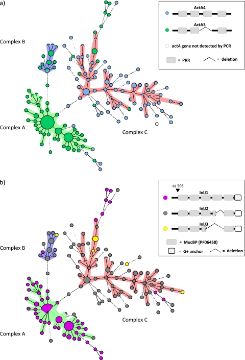
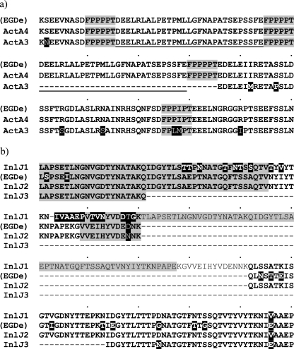
Listeria monocytogenes is among the most important food-borne pathogens and is well adapted to persist in the environment. To gain insight into the genetic relatedness and potential virulence of L. monocytogenes strains causing central nervous system (CNS) infections, we used multilocus variable-number tandem-repeat analysis (MLVA) to subtype 183 L. monocytogenes isolates, most from ruminant rhombencephalitis and some from human patients, food, and the environment. Allelic-profile-based comparisons grouped L. monocytogenes strains mainly into three clonal complexes and linked single-locus variants (SLVs). Clonal complex A essentially consisted of isolates from human and ruminant brain samples. All but one rhombencephalitis isolate from cattle were located in clonal complex A. In contrast, food and environmental isolates mainly clustered into clonal complex C, and none was classified as clonal complex A. Isolates of the two main clonal complexes (A and C) obtained by MLVA were analyzed by PCR for the presence of 11 virulence-associated genes (prfA, actA, inlA, inlB, inlC, inlD, inlE, inlF, inlG, inlJ, and inlC2H). Virulence gene analysis revealed significant differences in the actA, inlF, inlG, and inlJ allelic profiles between clinical isolates (complex A) and nonclinical isolates (complex C). The association of particular alleles of actA, inlF, and newly described alleles of inlJ with isolates from CNS infections (particularly rhombencephalitis) suggests that these virulence genes participate in neurovirulence of L. monocytogenes. The overall absence of inlG in clinical complex A and its presence in complex C isolates suggests that the InlG protein is more relevant for the survival of L. monocytogenes in the environment.
Figures
Similar articles
-
Increased spread and replication efficiency of Listeria monocytogenes in organotypic brain-slices is related to multilocus variable number of tandem repeat analysis (MLVA) complex.BMC Microbiol. 2015 Jul 3;15:134. doi: 10.1186/s12866-015-0454-0. BMC Microbiol. 2015. PMID: 26138984 Free PMC article.
-
Listeria monocytogenes sequence type 1 is predominant in ruminant rhombencephalitis.Sci Rep. 2016 Nov 16;6:36419. doi: 10.1038/srep36419. Sci Rep. 2016. PMID: 27848981 Free PMC article.
-
Ruminant rhombencephalitis-associated Listeria monocytogenes strains constitute a genetically homogeneous group related to human outbreak strains.Appl Environ Microbiol. 2013 May;79(9):3059-66. doi: 10.1128/AEM.00219-13. Epub 2013 Mar 1. Appl Environ Microbiol. 2013. PMID: 23455337 Free PMC article.
-
Listeria monocytogenes lineages: Genomics, evolution, ecology, and phenotypic characteristics.Int J Med Microbiol. 2011 Feb;301(2):79-96. doi: 10.1016/j.ijmm.2010.05.002. Epub 2010 Aug 13. Int J Med Microbiol. 2011. PMID: 20708964 Review.
-
Rhombencephalitis due to Listeria monocytogenes infection with GQ1b antibody positivity and multiple intracranial hemorrhage: a case report and literature review.J Int Med Res. 2021 Apr;49(4):300060521998568. doi: 10.1177/0300060521998568. J Int Med Res. 2021. PMID: 33866842 Free PMC article. Review.
Cited by
-
Large-Scale Comparison of Toxin and Antitoxins in Listeria monocytogenes.Toxins (Basel). 2020 Jan 2;12(1):29. doi: 10.3390/toxins12010029. Toxins (Basel). 2020. PMID: 31906535 Free PMC article.
-
Virulence Pattern Analysis of Three Listeria monocytogenes Lineage I Epidemic Strains with Distinct Outbreak Histories.Microorganisms. 2021 Aug 16;9(8):1745. doi: 10.3390/microorganisms9081745. Microorganisms. 2021. PMID: 34442824 Free PMC article.
-
Neurotropic Lineage III Strains of Listeria monocytogenes Disseminate to the Brain without Reaching High Titer in the Blood.mSphere. 2020 Sep 16;5(5):e00871-20. doi: 10.1128/mSphere.00871-20. mSphere. 2020. PMID: 32938704 Free PMC article.
-
Optimized Multilocus variable-number tandem-repeat analysis assay and its complementarity with pulsed-field gel electrophoresis and multilocus sequence typing for Listeria monocytogenes clone identification and surveillance.J Clin Microbiol. 2013 Jun;51(6):1868-80. doi: 10.1128/JCM.00606-13. Epub 2013 Apr 10. J Clin Microbiol. 2013. PMID: 23576539 Free PMC article.
-
Preliminary Investigation on Multiple-Locus Variable Number Tandem Repeat Analysis Profiles of Listeria Monocytogenes Isolates from Pork Meat Tested from Packaging to Fork.Ital J Food Saf. 2014 Jan 21;3(1):1660. doi: 10.4081/ijfs.2014.1660. eCollection 2014 Jan 21. Ital J Food Saf. 2014. PMID: 27800312 Free PMC article. Italian.
References
-
- Bartt R. 2000. Listeria and atypical presentations of Listeria in the central nervous system. Semin. Neurol. 20: 361–373 - PubMed
-
- Bibb W. F., Schwartz B., Gellin B. G., Plikaytis B. D., Weaver R. E. 1989. Analysis of Listeria monocytogenes by multilocus enzyme electrophoresis and application of the method to epidemiologic investigations. Int. J. Food Microbiol. 8: 233–239 - PubMed
-
- Borucki M. K., et al. 2004. Dairy farm reservoir of Listeria monocytogenes sporadic and epidemic strains. J. Food Prot. 67: 2496–2499 - PubMed
Publication types
MeSH terms
Substances
Associated data
- Actions
- Actions
- Actions
- Actions
- Actions
LinkOut - more resources
Full Text Sources
Medical
