

In vivo targeting of HER2-positive tumor using 2-helix affibody molecules
- PMID: 21984380
- PMCID: PMC4172459
- DOI: 10.1007/s00726-011-1096-7
In vivo targeting of HER2-positive tumor using 2-helix affibody molecules
Abstract
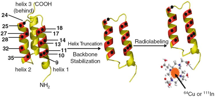
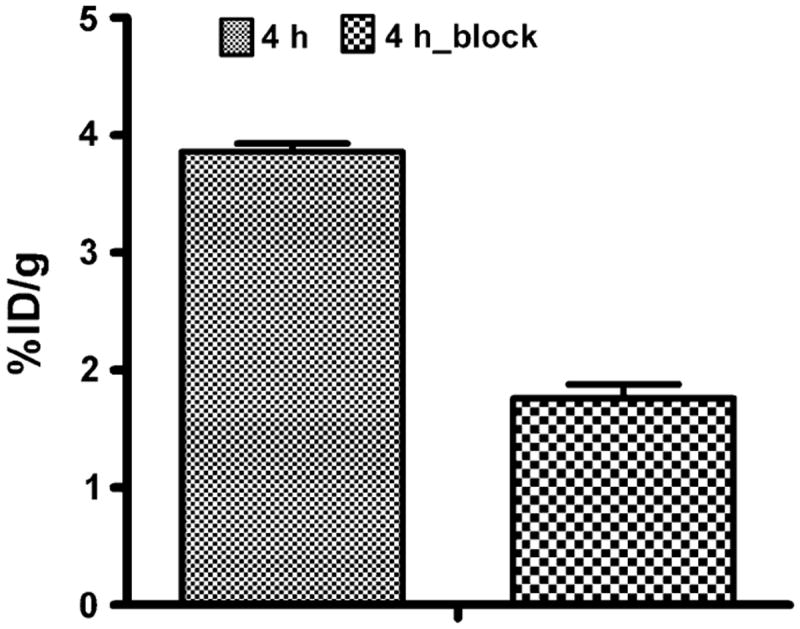
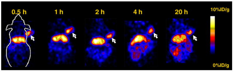
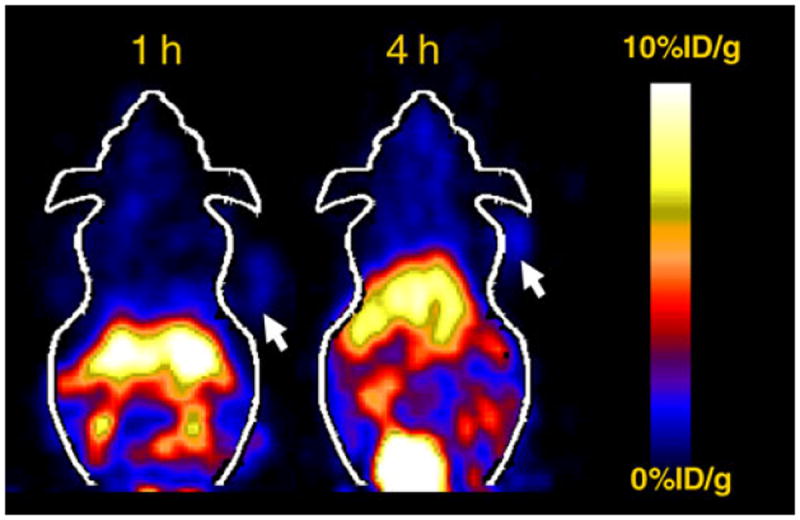
Molecular imaging of human epidermal growth factor receptor type 2 (HER2) expression has drawn significant attention because of the unique role of the HER2 gene in diagnosis, therapy and prognosis of human breast cancer. In our previous research, a novel cyclic 2-helix small protein, MUT-DS, was discovered as an anti-HER2 Affibody analog with high affinity through rational protein design and engineering. MUT-DS was then evaluated for positron emission tomography (PET) of HER2-positive tumor by labeling with two radionuclides, 68Ga and 18F, with relatively short half-life (t1/2<2 h). In order to fully study the in vivo behavior of 2-helix small protein and demonstrate that it could be a robust platform for labeling with a variety of radionuclides for different applications, in this study, MUT-DS was further radiolabeled with 64Cu or 111In and evaluated for in vivo targeting of HER2-positive tumor in mice. Design 1,4,7,10-tetraazacyclododecane-1,4,7,10-tetraacetic acid (DOTA) conjugated MUT-DS (DOTA-MUT-DS) was chemically synthesized using solid phase peptide synthesizer and I2 oxidation. DOTA-MUT-DS was then radiolabeled with 64Cu or 111In to prepare the HER2 imaging probe (64Cu/111In-DOTA-MUT-DS). Both biodistribution and microPET imaging of the probe were evaluated in nude mice bearing subcutaneous HER2-positive SKOV3 tumors. DOTA-MUT-DS could be successfully synthesized and radiolabeled with 64Cu or 111In. Biodistribution study showed that tumor uptake value of 64Cu or 111In-labeled DOTA-MUT-DS was 4.66±0.38 or 2.17±0.15%ID/g, respectively, in nude mice bearing SKOV3 xenografts (n=3) at 1 h post-injection (p.i.). Tumor-to-blood and tumor-to-muscle ratios for 64Cu-DOTA-MUT-DS were attained to be 3.05 and 3.48 at 1 h p.i., respectively, while for 111In-DOTA-MUT-DS, they were 2.04 and 3.19, respectively. Co-injection of the cold Affibody molecule ZHER2:342 with 64Cu-DOTA-MUT-DS specifically reduced the SKOV3 tumor uptake of the probe by 48%. 111In-DOTA-MUT-DS displayed lower liver uptake at all the time points investigated and higher tumor to blood ratios at 4 and 20 h p.i., when compared with 64Cu-DOTA-MUT-DS. This study demonstrates that the 2-helix protein based probes, 64Cu/111In DOTA-MUT-DS, are promising molecular probes for imaging HER2-positive tumor. Two-helix small protein scaffold holds great promise as a novel and robust platform for imaging and therapy applications.
Figures
References
-
- Ahlgren S, Wallberg H, Tran TA, Widstrom C, Hjertman M, Abrahmsen L, et al. Targeting of HER2-expressing tumors with a site-specifically 99mTc-labeled recombinant affibody molecule, ZHER2:2395, with C-terminally engineered cysteine. J Nucl Med. 2009;50:781–789. - PubMed
-
- Boswell CA, Sun X, Niu W, Weisman GR, Wong EH, Rheingold AL, et al. Comparative in vivo stability of copper-64-labeled cross-bridged and conventional tetraazamacrocyclic complexes. J Med Chem. 2004;47:1465–1474. - PubMed
-
- Capello A, Krenning EP, Breeman WA, Bernard BF, de Jong M. Peptide receptor radionuclide therapy in vitro using [111In-DTPA0]octreotide. J Nucl Med. 2003;44:98–104. - PubMed
Publication types
MeSH terms
Substances
Grants and funding
LinkOut - more resources
Full Text Sources
Other Literature Sources
Medical
Research Materials
Miscellaneous
